Japanese
English
- 有料閲覧
PPV購入案内
1,320円 (1,200円+税10%) こちらは、17ページから26ページの文献です。購入には医書.jp本会員登録が必要です。
会員登録完了後に改めて上記「購入サイトへ」を選択してください。
または「購入サイトへ」選択後に「購入ログイン」、「新規会員登録」の順に進んでください。
会員登録について詳しくはをご確認ください。
- Abstract 文献概要
- 1ページ目 Look Inside
抄録 ラット胎生延髄組織を同系ラット成体小脳内に移植し4〜9カ月間生存させた後,脳切片を作製し電顕および光顕的に観察した。延髄内の神経細胞の一部は小脳を標的組織としているが,この両組織間で移植実験を行い移植片内部および宿主・移植片境界域において,神経細胞,神経突起,シナプス形成,星状神経膠細胞(astroglia)や稀突起膠細胞(oligodendroglia)の増殖などの動態について形態学的に研究することを目的とした。本研究により以下の結果が得られた。1)移植片内部には,神経細胞の集団化や線維束の形成が観察され,部分的には延髄様構造への分化が認められた。神経細胞内の微小器官の発達は成熟型を示し,シナプス形成や髄鞘形成の像が観察された。2)宿主・移植片境界域には,無髄神経を主体とした神経突起が観察され,それらはastrogliaの増殖に伴って移植片から宿主内に侵入するものと考えられた。3)oilgodendrogliaの活性化を示唆する像が認められたが,有髄神経線維の宿主内侵入を観察することは非常に稀であった。
Pieces of medulla oblongata anlagen were dis-sected free from embryonic 13-20 day (E 13 to E 20) rat brain, and these were transplanted into the cerebellar vermis of adult rats (Fischer 344). After grafting, host animals survived for 4-9 months. Cytoarchitectonic organization of the graft and the relationship between host and graft were analyzed light microscopically in 34 animals using the Nissl and silver impregnation methods. Fine structures of the graft were analyzed in 4 animals using electron microscope.
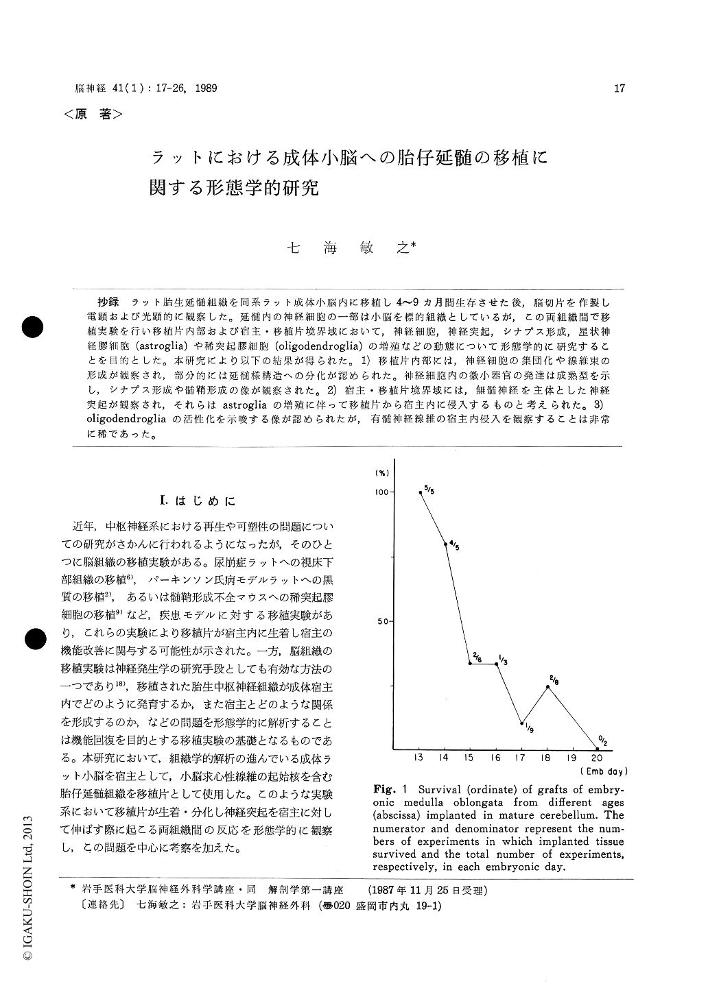
Grafts from E 13-14 donor tissue showed the highest survival rate (90%), which decreased as the donor embryonic age increased (i. e., E 15-16 : 33%, E 17-20 : 15%). In the surviving grafts, small (5-10 pm diameter), medium-sized (10-20 μm) and large (20-30 μm) neurons, whose cytoplasmic organelles appeared normal, were observed. Bundles of myelinated fibers traversed in every direction and neurons were often clustered, indica-ting characteristic features of the medulla ob-longata. Electron microscopically, various types of synaptic formations were also observed. De-generative profiles of nerve-fiber endings, contain-ing dense bodies and lysosomic figures, were also seen. The degeneration seemed to be caused by the failure of their establishing connections with their proper targets in the host.
In both the host tissue and the graft-host inter-face, neuronal processes apparently derived from the graft were frequently observed. Some axonal processes contained large-cored vesicles, and some dendritic processes were enlarged at their stalks and tips. Aberrant axon terminals of unmyelinated fibers in the host medullary layer were considered to be the graft origin. These fibers were always accompanied by prominent glial proliferation. There was no indication of forming myelinated fiber bundles that entered the host cerebellum from the donor tissue, although the former wasthe target of the latter. Cell bodies of host granule cells and oligodendroglia in the graft-host interface were surrounded by procceses of oli-godendroglia, forming thin myelin lamellae.
The present study showed that unmyelinated fibers from the graft could enter the host cerebel-lum non-specifically accompanied by glial prolifera-tion, whereas myelinated fibers could hardly penetrate into the host crossing the glial scar.
Copyright © 1989, Igaku-Shoin Ltd. All rights reserved.