Japanese
English
- 有料閲覧
PPV購入案内
1,320円 (1,200円+税10%) こちらは、609ページから615ページの文献です。購入には医書.jp本会員登録が必要です。
会員登録完了後に改めて上記「購入サイトへ」を選択してください。
または「購入サイトへ」選択後に「購入ログイン」、「新規会員登録」の順に進んでください。
会員登録について詳しくはをご確認ください。
- Abstract 文献概要
- 1ページ目 Look Inside
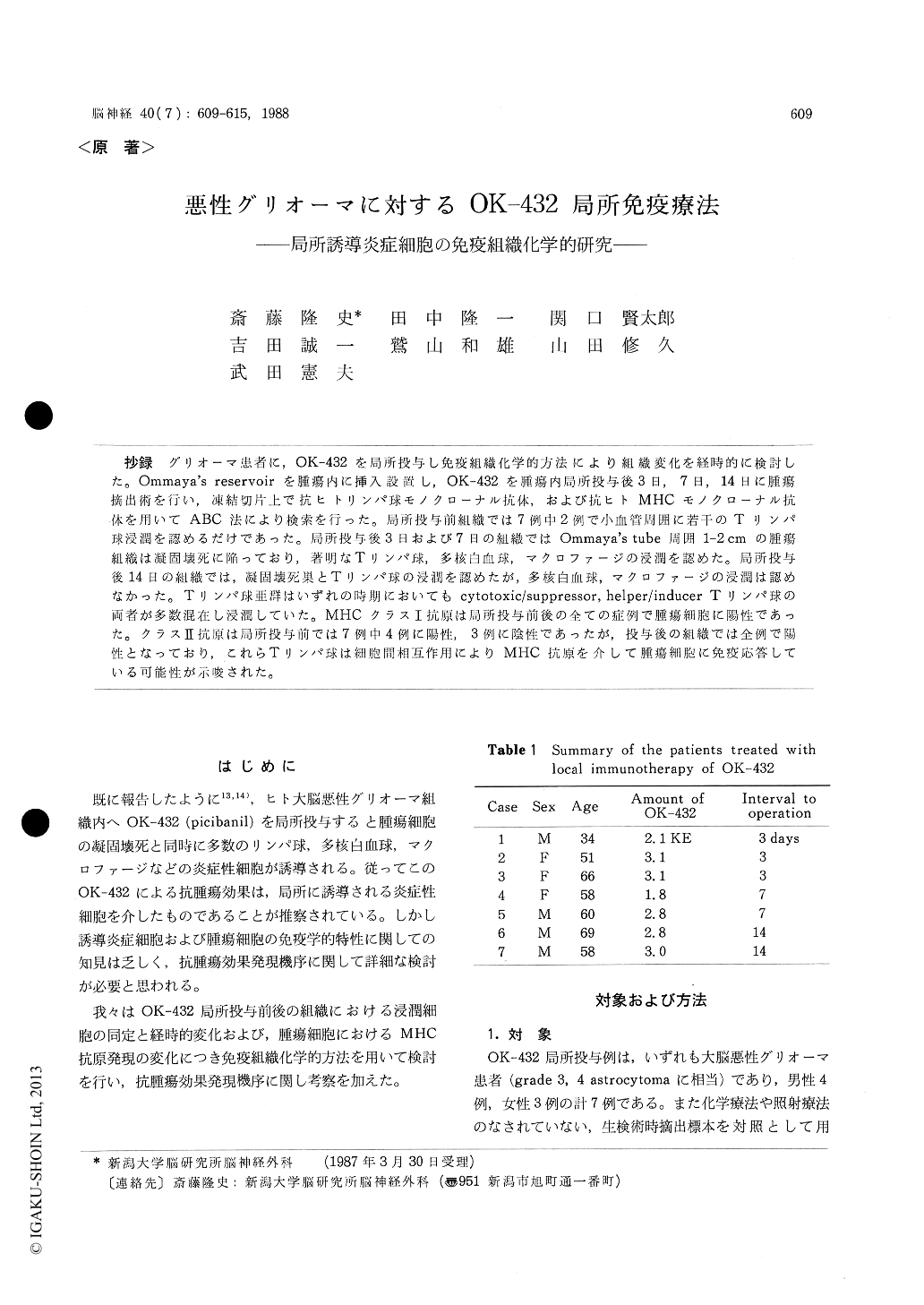
抄録 グリオーマ患者に,OK−432を局所投与し免疫組織化学的方法により組織変化を経時的に検討した。Ommaya's reservoirを腫瘍内に挿入設置し,OK−432を腫瘍内局所投与後3日,7日,14日に腫瘍摘出術を行い,凍結切片上で抗ヒトリンパ球モノクローナル抗体,および抗ヒトMHCモノクローナル抗体を用いてABC法により検索を行った。局所投与前組織では7例中2例で小血管周囲に若干のTリンパ球浸潤を認めるだけであった。局所投与後3日および7日の組織ではOmmaya's tube周囲1-2cmの腫瘍組織は凝固壊死に陥っており,著明なTリンパ球,多核白血球,マクロファージの浸澗を認めた。局所投与後14日の組織では,凝固壊死巣とTリンパ球の浸潤を認めたが,多核白血球,マクロファージの浸潤は認めなかった。Tリンパ球亜群はいずれの時期においてもcytotoxic/suppressor, helper/inducer Tリンパ球の両者が多数混在し浸潤していた。MHCクラスI抗原は局所投与前後の全ての症例で腫瘍細胞に陽性であった。クラスII抗原は局所投与前では7例中4例に陽性,3例に陰性であったが,投与後の組織では全例で陽性となっており,これらTリンパ球は細胞間相互作用によりMHC抗原を介して腫瘍細胞に免疫応答している可能性が示唆された。
Chronological changes of glioma tissues treated with local immunotherapy with OK-432 were ex-amined by immunohistochemical method. OK-432 was injected into glioma tissues through Ommaya's reservoir 3 days (3 patients), 7 days (2 patients) and 14 days (2 patients) prior to the operation. Frozen sections surgically obtained from these patients were stained with avidin-biotin-peroxidase com-pex method using Leu-series monoclonal anti-bodies for pan T lymphocytes (Leu-1), cytotoxic/ suppressor T lymphocytes (Leu-2 a), helper/inducer T lymphocytes (Leu-3 a), B lymphocytes (Leu-12), MHC class I antigen (β2m) and MHC class II antigen (HLA-DR).
In 2 out of 7 glioma tissues obtained before local injection of OK-432, only few T lymphocytes were found infiltrating around the small blood vessels. In all glioma tissues obtained 3 and 7 days after injection, coagulation necrosis of glioma tissues was observed within 1-2 cm from Ommaya's tubeand many T lymphocytes granulocytes and macro-phages were infiltrating diffusely in the glioma tissues. Whereas in all glioma tissues obtained 14 days after injection, coagulation necrosis was also observed, however granulocytes and macrophages were scares. The most of the infiltrating cells were T lymphocytes. Examination of T lympho-cytes phenotypes revealed that both cytotoxic/sup-pressor and helper/inducer phenotypes of T lym-phocytes were intermingled with each other in all cases.
β2m) was expressed on the most of glioma cells in all cases before and after injection. Whereas HLA-DR antigen was expressed on the tumor cells in 4 out of 7 cases before injection, however this antigen was expressed in all cases after injection. These results suggest that both phenotypes of T lymphocytes react to the tumor cells through T lymphocytes interaction with MHC antigens on the tumor cells.
Copyright © 1988, Igaku-Shoin Ltd. All rights reserved.