Japanese
English
- 有料閲覧
- Abstract 文献概要
- 1ページ目 Look Inside
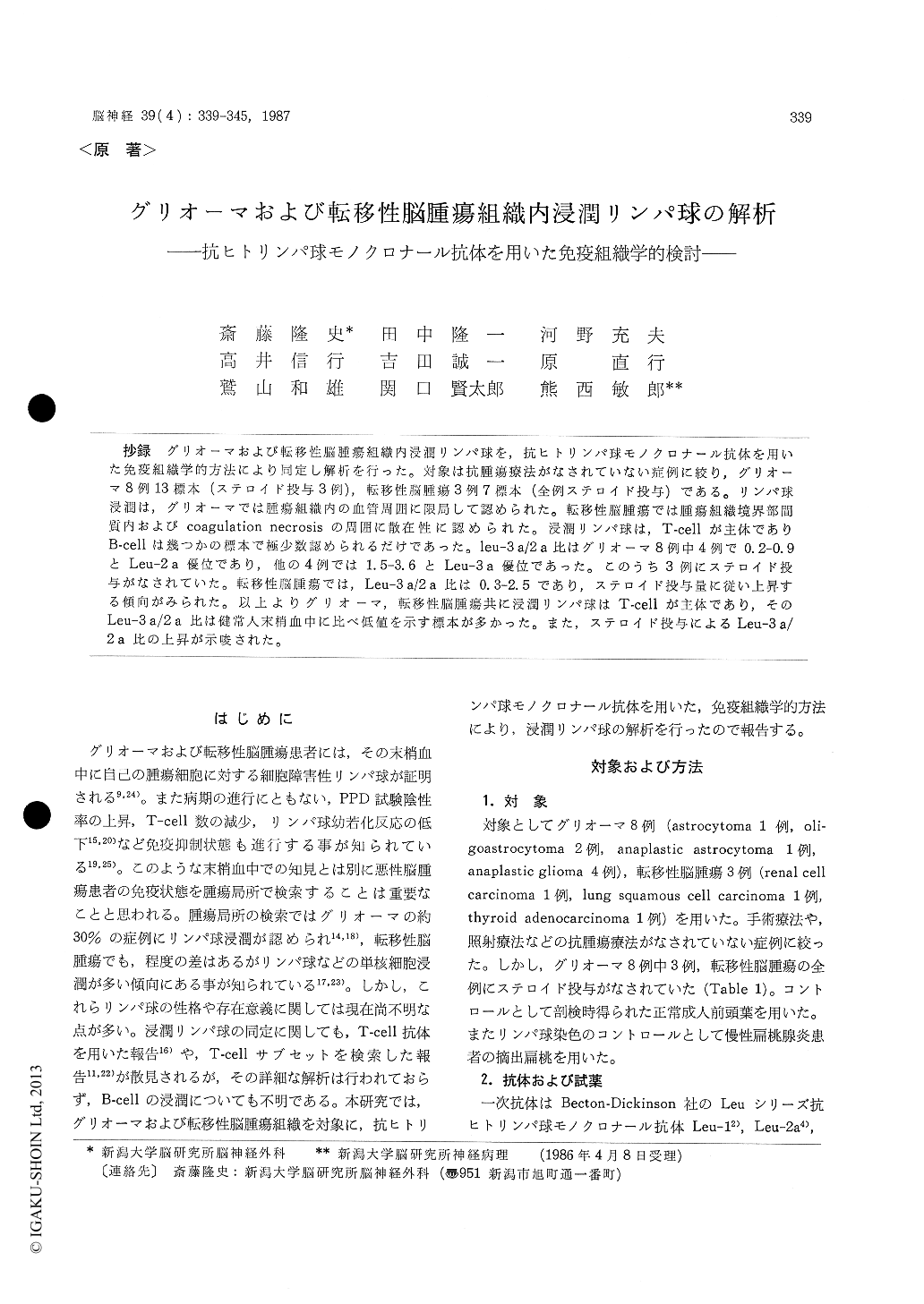
抄録 グリオーマおよび転移性脳腫瘍組織内浸潤リンパ球を,抗ヒトリンパ球モノクロナール抗体を用いた免疫組織学的方法により同定し解析を行った。対象は抗腫瘍療法がなされていない症例に絞り,グリオーマ8例13標本(ステロイド投与3例),転移性脳腫瘍3例7標本(全例ステロイド投与)である。リンパ球浸潤は,グリオーマでは腫瘍組織内の血管周囲に限局して認められた。転移性脳腫瘍では腫瘍組織境界部間質内およびcoagulation necrosisの周囲に散在性に認められた。浸潤リンパ球は,T-cellが主体でありB-cellは幾つかの標本で極少数認められるだけであった。leu−3a/2a比はグリオーマ8例中4例で0.2味0.9とLeu−2a優位であり,他の4例では1.5-3.6とLeu−3a優位であった。このうち3例にステロイド投与がなされていた。転移性脳腫瘍では,Leu−3a/2a比は0.3-2.5であり,ステロイド投与量に従い上昇する傾向がみられた。以上よりグリオーマ,転移性脳腫瘍共に浸潤リンパ球はT-cellが主体であり,そのLeu−3 a/2 a比は健常人末梢血中に比べ低値を示す標本多かった。また,ステロイド投与によるLeu−3a/2a比の上昇が示唆された。
Subpopulations of infiltrating lymphocytes were studied by immunohistological method using monoclonal antibodies in gliomas and metastatic brain tumors. Thirteen specimens from 8 glioma patients, and 7 specimens from 3 metastatic brain tumor patients were used. No special therapy for brain tumor had been performed in these cases, but 3 glioma patients and all metastatic brain tumor patients had received steroid hormone. Frontal lobe obtained from the autopsy case of chest trauma was served as a normal control. Frozen sections were stained with avidin-biotinperoxidase complex method using Leu-series monoclonal antibodies for pan T-cells (Leu-1) , cytotoxic/suppressor T-cells (Leu-2 a) , helper/ inducer T-cells (Leu-3 a) and B-cells (Leu-12). Lymphocyte infiltrates were quantitated by counting positively stained cells in 13 glioma and 7 metastatic brain tumor specimens. In normal frontal lobe, only a few T-cells infiltrated around several blood vessels in the parenchyma and subarachnoid space. But in the cases of glioma, many perivascular lymphocytic infiltrates were found and in the cases of metastatic brain tumor, many lymphocytes were found diffusely in the interstitial area between nests of tumor cells. Most of these lymphocytes were T-cells and B-cells were scarce, and Leu-2 a and Leu-3 a positive cells intermingled with each other. Len-3 a/2 a ratio ranged from O. 2 to O. 9 in the half of gliomas and 1.5 to 3. 6 in another half of gliomas, three of which were treated with ste-roid hormone. In the cases of metastatic brain tumor this ratio ranged from 0.3 to 2.5 and higher values were observed in cases which had received higher doses of steroid hormone.

Copyright © 1987, Igaku-Shoin Ltd. All rights reserved.


