Japanese
English
- 有料閲覧
PPV購入案内
1,320円 (1,200円+税10%) こちらは、281ページから287ページの文献です。購入には医書.jp本会員登録が必要です。
会員登録完了後に改めて上記「購入サイトへ」を選択してください。
または「購入サイトへ」選択後に「購入ログイン」、「新規会員登録」の順に進んでください。
会員登録について詳しくはをご確認ください。
- Abstract 文献概要
- 1ページ目 Look Inside
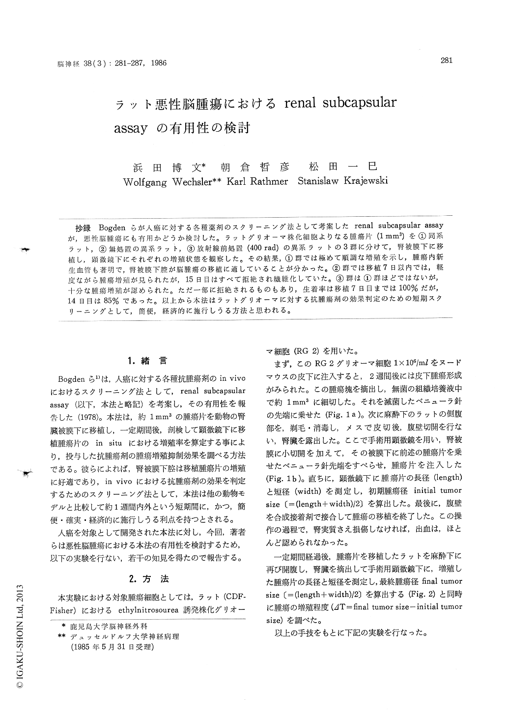
抄録 Bogdenらが人癌に対する各種薬剤のスクリーニング法として考案したrenal subcapsular assayが,悪性脳腫瘍にも有用かどうか検討した。ラットグリオーマ株化細胞よりなる腫瘍片(1mm3))を①同系ラット,②無処置の異系ラット,③放射線前処置(400rad)の異系ラットの3群に分けて,腎被膜下に移植し,顕微鏡下にそれぞれの増殖状態を観察した。その結果,①群では極めて順調な増殖を示し,腫瘍内新生血管も著明で,腎被膜下腔が脳腫瘍の移植に適していることが分かった。②群では移植7日以内では,軽度ながら腫瘍増殖が見られたが,15日目はすべて拒絶され繊維化していた。③群は①群ほどではないが,十分な腫瘍増殖が認められた。ただ一部に拒絶されるものもあり,生着率は移植7日目までは100%だが,14日目は85%であった。以上から本法はラットグリオーマに対する抗腫瘍剤の効果判定のための短期スクリーニングとして,簡便,経済的に施行しうる方法と思われる。
As a rapid screening method for testing chemotherapeutic agents against human tumor explants, renal subcapsular assay has been established by Bogden et al (1978).
The authors investigated the effectiveness of the technique of Bogden for rat glioma.
Preselected 1mm3) fragments of viable tumor tissue of rat (CDF) were implanted under the renal capsule of normal immunocompetent rats. Then initial and final tumor sizes ((length+width)/2) were measured in situ with an oculometer inserted into a surgical microscope, and evaluated the changes of tumor sizes.
The experiments were divided into three groups. The first group was that tumor pieces of rat (CDF) were implanted under the renal capsule of the syngenetic rats (CDF). Non-irradiated and preirradiated (400 rad) heterogenetic rats (Wistar) were used as the second and third groups.
The explanted tumor pieces grew rapidly in the first group (syngenetic rats). On the other hand, poor growth of the second group (non-irradiated heterogenetic rats) was seen within 7 days after implantation, and rejected completely until 15 days after implantation. Explanted tumor pieces in the third group (pre-irradiated heterogenetic rats), however, grew enough big, though the change of tumor sizes was less than that of the first group.
Histological examination was also performed. In the first and third group, proliferation of glioma cells was remarkable under the renal capsule, and neovascularization in the explanted tumor tissue was provided from the capsule, first, and then parenchym of the kidney. Even necrosis and hemorrhage were seen in the tumor tissue 15 days after implantation.
In the second group, growth of tumor cells was poor mixed with reactive infiltration of lymphcells and fibroblasts. Finally, 15 days after implantation, only fibrosis was observed under the renal capsule.
Anyway, it was possible to find good enough growth of the explanted tumor pieces by using the syngenetic rats or immunosuppressive method (pre-irradiation) in renal subcapsular assay.
In conclusion, renal subcapsular assay for rat glioma proved to be a convenient, rapid and economical technique as a screening method for testing chemotherapeutic agents.
Copyright © 1986, Igaku-Shoin Ltd. All rights reserved.