Japanese
English
- 有料閲覧
- Abstract 文献概要
- 1ページ目 Look Inside
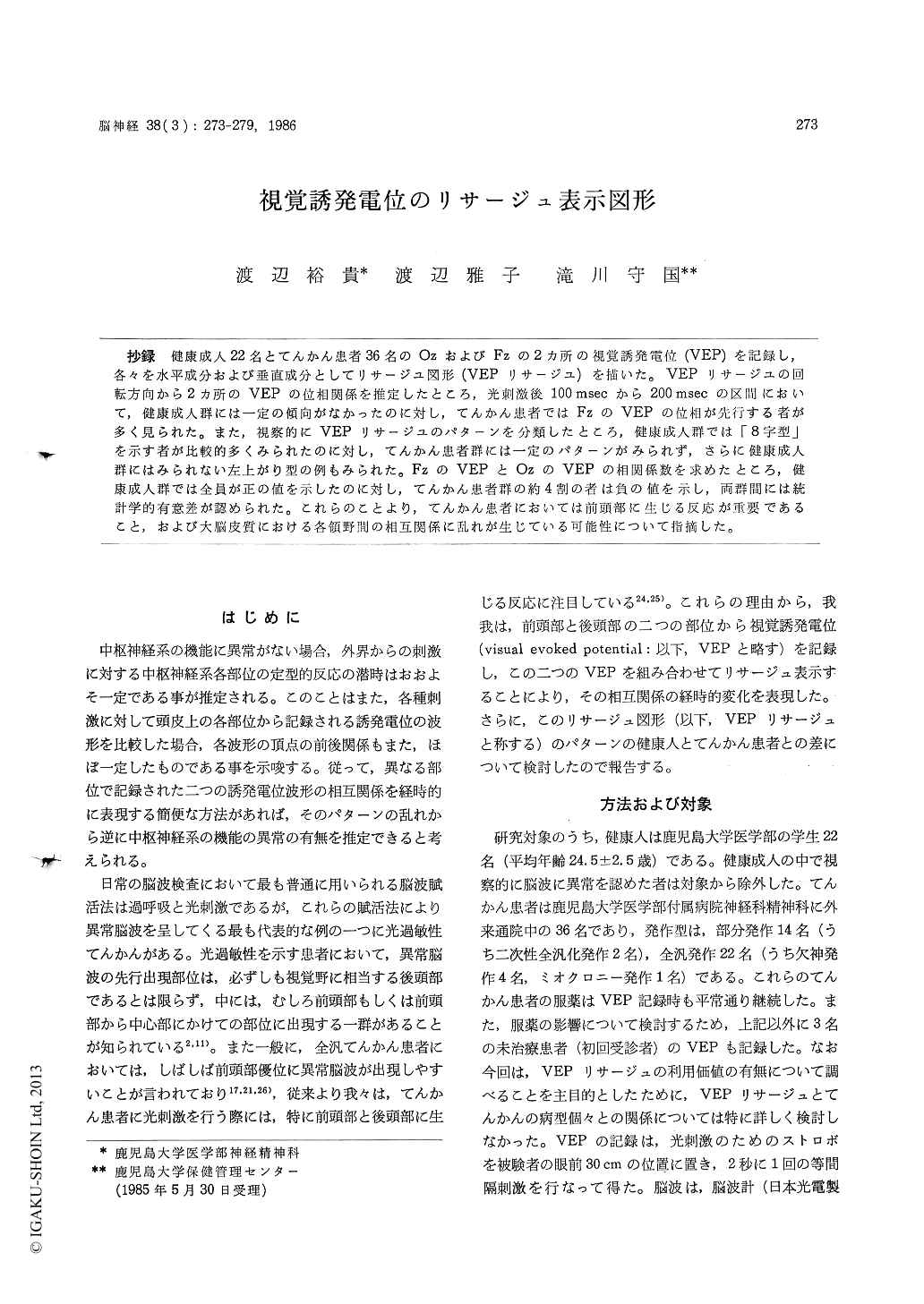
抄録 健康成人22名とてんかん患者36名のOzおよびFzの2カ所の視覚誘発電位(VEP)を記録し,各々を水平成分および垂直成分としてリサージユ図形(VEPリサージユ)を描いた。 VEPリサ—ジユの回転方向から2カ所のVEPの位相関係を推定したところ,光刺激後100 msecから200 msecの区間において,健康成人群には一定の傾向がなかったのに対し,てんかん患者ではFzのVEPの位相が先行する者が多く見られた。また,視察的にVEPリサージユのパターンを分類したところ,健康成人群では「8字型」を示す者が比較的多くみられたのに対し,てんかん患者群には一定のパターンがみられず,さらに健康成人群にはみられない左上がり型の例もみられた。FzのVEPとOzのVEPの相関係数を求めたところ,健康成人群では全員が正の値を示したのに対し,てんかん患者群の約4割の者は負の値を示し,両群間には統計学的有意差が認められた。これらのことより,てんかん患者においては前頭部に生じる反応が重要であること,および大脳皮質における各領野間の相互関係に乱れが生じている可能性について指摘した。
The value of the visual evoked potential (VEP) technique for diagnosis of epilepsy is limited. Except for photosensitive epilepsies, the visual evoked potentials (VEPs) do not reveal more than a routine EEG. As one of the reasons of that, it is pointed out that a review of VEP literature reveals different "normal values" for peak latencies and amplitudes. Thus in this study, lissajous figures of VEPs (VEP-L) were attempted instead of the measurement of peak latencies and amplitudes of VEPs. Thirty six epileptic subjects (14 have partial seizures, 22 have gene-ralized seizures) and 22 control subjects were in-vestigated. Several EEGs to storoboscopic flashes were recorded from scalp electrodes placed at the frontal (Fz) and occipital (Oz) regions ac-cording to the 10-20 electrode system, using a right ear as nonreference with a EEG amplifier (Nihon Kohden ME-175 E) and a data recorder (TEAC R-60). Then the phasic relationships bet-ween the VEPs from the two areas were analyzedwith a medical computer (Nohon Kohden ATAC-2300) and printed out as VEP-L.
The results are summerized as follows : First, as time passes from 100 msec to 200 msec in the VEP-L, the epileptic patient group showed more right-rated types than that of the control group (p<0.01).
Second, the VEP-L at the periods from 50 msec after photic stimulus, were classified into 5 types inspectively. The 5 types are the right-ascending type, the left-ascending type, the vertical type, the horizontal type, and the circular type. In control subjects, a type of the VEP-L that is the right-ascending, simple type like 8-lettering was observed most frequently (about 45%), and the left-ascending type was never observed. In cont-rast, in epileptic patients, all of the 5 types were observed, and almost of patterns of the VEP-Lwere complex, and there was no dominant type of the VEP-L.
Finally, correlation coefficients (R) between Oz-VEP and Fz-VEP, which reveals one of quantita-tive aspeces of the VEP-L, were calculated with a medical computer. All control subjects showed positive R only. But epileptic patienst showed both positive and negative Rs. The difference between values of the two groups are statistically significant (p <0.05).
It is suggested that in the VEPs which are recorded from epileptic patients, Fz-VEP may be more important than other VEPs for clinical use, and that inter-cortical relationships in the epilepsy may be confused. And it is suggested that the VEP-L technique is useful for the analy-sis of VEPs in epileptic patients.

Copyright © 1986, Igaku-Shoin Ltd. All rights reserved.


