Japanese
English
- 有料閲覧
- Abstract 文献概要
- 1ページ目 Look Inside
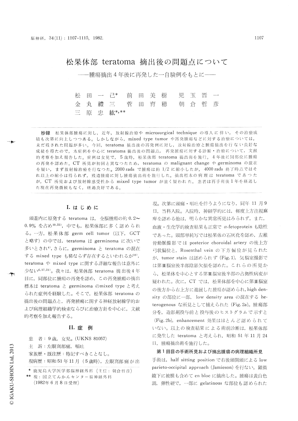
抄録 松果体部腫瘍に対し,近年,放射線治療やmicrosurgical techniqueの導入に伴い,その治療成績も次第に向上しつつある。しかしながら,mixed type tumorや再発腫瘍などに対する治療については,未だ残された問題が多い。今回,teratoma摘出後の在発例に対し,放射線治療と腫瘍摘出を行ない良好な成績を得たので,本症例を中心にteratoma摘出後の問題点、再発腫瘍に対する診断・治療について,文献的考察を加え報告した。症例は女児で,5歳時,松果体部teratoma摘出術を施行。4年後に同部位に腫瘍の再発を認めた。CT所見が初回と異なつたため,teratomaのmalignant changeやgerminomaの混在を疑い,まず放射線治療を行なつた。2000radsで腫瘍は約1/2に縮小したが,4000 rads終了時点ではそれ以上の縮小は得られず,残遺腫瘍に対し腫瘍摘出術を施行した。摘出標本の病現はteratomaであつたが,CT所見および放射線感受性からmixed type tumorが強く疑われた。患者は再手術後1年を経過した現在再発徴候もなく,経過良好である。
A case of teratoma in the pineal region which recurred 4 years after the first tumor removal was reported in this paper.
When the patient was 5 years old, she, complain-ed of headache and vomiting, and visited our hospital. As a heterogeneous mass with no enhan-cement effect was found in the pineal region by CT scan, she admitted on November 9, 1976. There was no abnomalities on physical examina-tion but neurological examination revealed slight disturbance of conjugate upward gaze (Parinaud's sign). Left vertebral angiogram demonstrated pos-terior superior displacement of posterior cho-roidal artery and downward displacement of Ro-senthal vein, but early venous filling and tumor stain were not seen. Under preoperative diagno-sis of a teratoma in the pineal region, the first operation (left occipital craniotomy and total re-moval of the tumor) was performed on November 24, 1975. Microscopic examinations revealed that the removed tumor was a mature teratoma in the pineal region.
Postoperative course was uneventful and dis-charged on December 20, 1975. The follow-up study was continued at outside clinic after dis-charge. There was no signs of recurrence until 3 years after the first operation, but on January, 1981 (4 years after the first operation), she suffered from severe headache and vomiting again and re-admit-ted to our hospital on February 3, 1981.
There was no remarkable neurological deficits except for the mild intracranial hypertensive sign and no changes of findings on angiogram. But CT findings were markedly characteristic. It revealed a heterogenous mass with remarkable enhancement effect in the pineal region and ventricular enlar-gement. Because a mixed type (teratomatous and germinomatous) of pineal tumors was suspected from the CT findings, irradiation was done after V-P shunt.
The tumor was reduced to half size after the first course of 2000 rads irradiation, but there is no more reduction of the size of the tumor follow-ing the second course of 2000 rads (total 4000 rads) ii radiation.
Against the residual tumor, tumor removal was performed on June 2, 1981. Micros,opically, the most part of the resected tumor showed fibrous changes caused by irradiation and partially tera-tomatous compartment.
From this result (radiosensitivity and histology) the authors assumed that the recurred tumor could be a mixed type (germinoma and teratoma) of pineal tumor.
Postoperative course was uneventful except for a transient disturbance of conjugate upward gaze and she discharged on June 25, 1981. And now, there is no signs of recurrence 12 months after the second operation.
Conclusively, it will be stressed that we should continue follow-up study the case even after total removal of teratoma, especially in the pineal re-gion. Moreover, it was considered that there is a posibility of the changes of the histological fea-tures on recurrence of the pineal teratoma. When germinomatous compartment is suspected, irradi-ation is the first choice and then microsurgical operation should be done against residual tumor.

Copyright © 1982, Igaku-Shoin Ltd. All rights reserved.


