Japanese
English
- 有料閲覧
PPV購入案内
1,320円 (1,200円+税10%) こちらは、201ページから205ページの文献です。購入には医書.jp本会員登録が必要です。
会員登録完了後に改めて上記「購入サイトへ」を選択してください。
または「購入サイトへ」選択後に「購入ログイン」、「新規会員登録」の順に進んでください。
会員登録について詳しくはをご確認ください。
- Abstract 文献概要
- 1ページ目 Look Inside
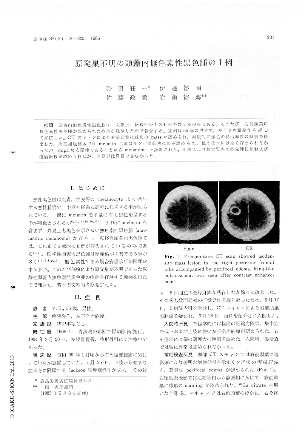
抄録 頭蓋内無色素性黒色腫霞は,文献上,転移性のもの6例を数えるのみである。このたび,右前頭葉に無色素性黒色腫が認められた症例を経験したので報告する。症例は55歳の男性で,左半身痙攣発作を起して来院した。CTスキャンにより右前頭葉に球形のmassが認められ,肉眼的に赤色の易出血性の腫瘍を摘出した。病理組織標本ではmelanin色素はリンパ節転移にのみ認められ,他の標本には全く認められなかったが,dopa反応陽性であることからmelanomaと診断された。剖検により脳実質内の多発性転移および遠隔転移が認められたが,原発巣は発見できなかった。
Malignant melanoma is usually black in color because of the existence of melanin pigments in it. Differing from such a usual melanoma, amela-notic melanoma, which has no melanin pigments and is not black-colored, is rarely described in literature. In this report, a case of intracranial amelanotic melanoma of unknown origin is pre-sented and discussed. This represents the seventh case report of an intracranial amelanotic melano-ma.
On May 28, 1984, a 55-year-old man was admit-ted to our department because of repeated left hemiconvulsion. Neurological and physical exami-nation disclosed left hemiparesis, poor concentra-tion and right inguinal tumors. CT scan revealed a right posterior frontal mass with ring like enhancement. On June 7, the patient underwent craniotomy. The brain tumor, partly emerged from right frontal lobe, was reddish and easy to bleed. Macroscopically there were no abnormalfindings over leptomeninges. The right inguial tumors, that were lymph nodes, were also re-moved simultaneously. Microscopic interpretation at that time was that of a malignant tumor of ecto-dermal origin. Although no melanin pigments were demonstrated in any specimens except for a part of lymph nodes, dopa reaction was positive in both specimens from the brain tumor and the inguial lymph nodes, strongly suggesting amela-notic melanoma. Postoperative course was unevent-ful, but CT scan of Aug. 31 again showed mul-tiple intracerebral enhanced lesion and ventricular hemorrhage. Aphasia, right hemiparesis and cons-ciousness disturbance developed gradually, and he died on Sept. 21, 1984. Autopsy demonstrated multiple intracerebral hematomas, containing much tumor cells in lung, thyroid and subcutaneous tissue. Primary lesion remained unknown in spite of an extensive examination in autopsy
Malignant melanoma is usually described to be black and to include various amounts of melanin pigments. Amelanotic melanoma, which has no melanin pigments, is rarely encountered, and in-tracranial amelanotic one is extremely rare. Only six cases of intracranial amelanotic melanoma have been reported in literature. The pathological diagnosis of intracranial amelanotic melanoma have been reported in literature. The pathological diagnosis of intracranial amelanotic melanoma of unknown origin is difficult and dopa reaction is very helpful.
Copyright © 1986, Igaku-Shoin Ltd. All rights reserved.