Japanese
English
- 有料閲覧
- Abstract 文献概要
- 1ページ目 Look Inside
I.はじめに
中枢神経系原発の黒色腫はきわめて稀な疾患ではあるが,文献上報告は少くない。たとえば,臨床的研究は,松本13),寺尾19),牧12),Piscol14)らにより,電顕的観察は,木下8),Ward21)らにより,化学療法は,Grooms4),Hill6),Beretta1)らにより詳しく報告されている。しかし,予後は一般に不良で,長期生存例はきわめて稀である。我々は本疾患で初回治療より12年間以上生存している症例を経験した。その長期生存の理由に関して,本腫瘍のDNA分析を行い,さらに治療にDTIC (dimethyltriazenoimidazole carbo xamide,以下DTICと略す)を使用したので,これらの意義に若干の考察を加えて報告する。
The patient, 46 years old : male, had congenital lentigo in upper middle abdominal skin.
A blue nav us, 12 × 16 cm in diameter at left tempoparietal region, and a pea-sized pigmentation on left conjunctiva bulbi was noted. There is nothing particular in his familial history.
At his 33 years of age, he was admitted in the Tokyo University Hospital with complainning of headache, vommitting, diplopia, and diminishing of right visual activity. An angiography revealed tumor in left middle fossa. In November, 1966, partial removal of the tumor was performed, and clinical diagnosis of a melanoma was established. Histologically, it was a melanoblastoma with rather less cellularity, consisted of spindle cells containing abundant melanin granule.
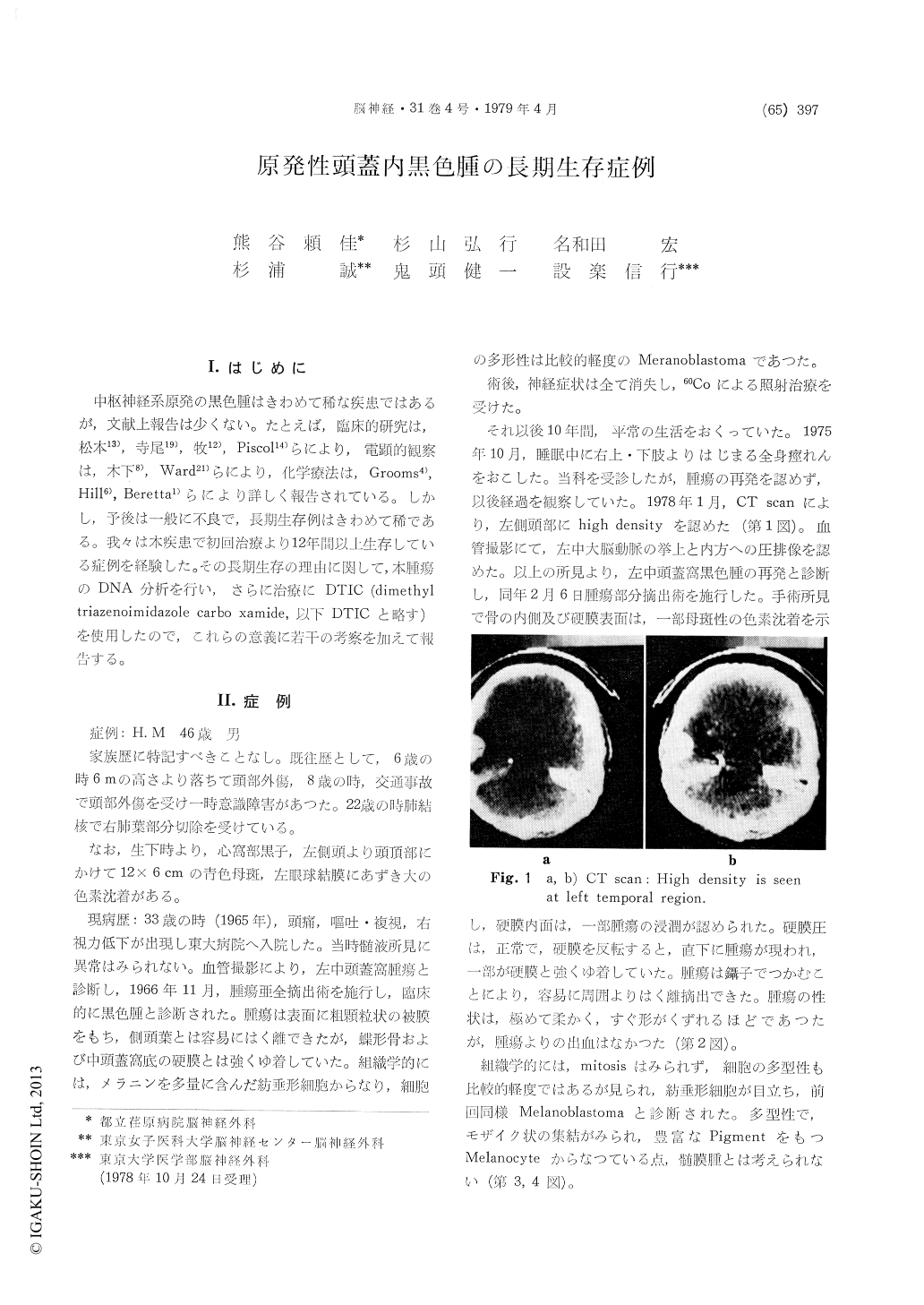
After operation, 60Co radiation therapy was added. Since them, he had had no complainds nor neuro-logical deficits during 10 years, and spent normal social life. In May 1975, general convulsive seizures originaling in right upper and low ex-tremities appeared. No recurrence of the tumor was noted at that time. But, in January 1978, CT and angiography revealed the recurrence of the tumor in left temperal region. On February 6, 1978, second partial removal of the tumor was performed.
The inner surface of skull and dura mater were invaded by tumor. The main part of the tumor was intradurally located and tightly adhesed to dura mater.
The soft tumor was easily separated from the surrounding tissue and removed out. No hemor-rhage was noted.
The tumor was packed with spindle cells. No mitoses nor irregularity of cellular or nuclear shapes were noted. The diagnosis of Melano-blastoma was reconfirmed.
Electron microscopic findings revealed its multi-plisity of cells. It was consisted of partly mosaic shaped cells and partly melanocytes with abundant pigment granules.
The chemotherapy with Vincristine (1 mg), ACNU (100 mg), DTIC (450 mg) was administed in ravenously. The above chemotherapy was repeated 3 times in every 4 weeks. The patient has been well afterward.
DNA analysis of this tumor cells was carried out by flow micro fluorometry. The ratio of the cells in S, G2 and M phase was smaller than that of metastatic intracranial melanoma from skin. This result revealed the longer doubling time of this tumor.
Copyright © 1979, Igaku-Shoin Ltd. All rights reserved.