Japanese
English
- 有料閲覧
PPV購入案内
1,320円 (1,200円+税10%) こちらは、1137ページから1142ページの文献です。購入には医書.jp本会員登録が必要です。
会員登録完了後に改めて上記「購入サイトへ」を選択してください。
または「購入サイトへ」選択後に「購入ログイン」、「新規会員登録」の順に進んでください。
会員登録について詳しくはをご確認ください。
- Abstract 文献概要
- 1ページ目 Look Inside
I.緒言
頭蓋内の正中部または傍正中部を走行する深部脳静脈の走行位置は正常変異が最も少なく,一定の形態を示し,頭蓋の形態とも密接な相関関係がある。従つて深部脳静脈は脳腫瘍の局在診断においてその診断的価値が一般に認められている。
従来主としてモンロー孔に相当する静脈角および極く一部に内大脳静脈の位置については計測法が種々考案され発表された。しかし従来の計測法はいずれも非常に複雑なものが多く,内大脳静脈,脳底静脈およびガレン大静脈からなる深部脳静脈全体の特有な形態を計測的に扱つた研究がみあたらない。
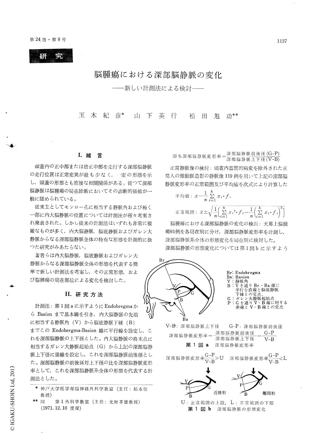
A new method of measurements of the configu-ration of the deep cerebral venous system, which is composed of the internal cerebral and basal veins, was devised. The ratio, antero-posterior distance to supero-inferior distance of the deep cerebral veins as shown in Fig. I, was considered to represent the whole configuration of the deep cerebral veins, and was named as deformity index of the deep cerebral veins.
After the mean values and the normal limits of this deformity index of the deep cerebral veins were established, the deformity index in the supra-tentorial tumors in various location was discussed. The following results could be obtained from present study.
The frontal tumors could be in most cases very satisfactorily localized by our measurements.
Tumors in the occipital, temporal and deep areas unless large enough to cause obvious changes in the deep cerebral veins, could not be constantly localized, therefore it must be always kept in mind that tumors in these location, if small, may not cause obvious deformity of the deep cerebral veins, and the measurements may sometimes give mislead-ing information.
It was pointed out that our method of measure-ments was of great importance in differetiationbetween extracerebral tumors and intracerebral tumors of the temporal region, since, the internal carebral veins and basal vein showed closed configu-ration in the extracerebral temperal tumors while both veins were normal or separated each other in the intracerebral tumors of the temporal lobe.
Our method of measurements was of great diag-nostic value in localizing the supratentorial space occuping lesions, since valuable informations as mentined above could be obtained from it, and changes of the whole configuration of the deep cerebral veins could be represented by it, and more-over the measurement was very simple and useful.
The authors, however, would like to point out in conclusion that we must not merely rely on the actual results of measurements, although it is of diagnostic value. The method of measurements has of course limitations in localizing the brain tumors and may give misleading informations, since the attempt is made to measure a three-dimensional displacement in a one dimensional plane, and there are also relatively wide range of normal variations of the deep cerebral veins.
Copyright © 1972, Igaku-Shoin Ltd. All rights reserved.