Japanese
English
- 有料閲覧
PPV購入案内
1,320円 (1,200円+税10%) こちらは、1239ページから1250ページの文献です。購入には医書.jp本会員登録が必要です。
会員登録完了後に改めて上記「購入サイトへ」を選択してください。
または「購入サイトへ」選択後に「購入ログイン」、「新規会員登録」の順に進んでください。
会員登録について詳しくはをご確認ください。
- Abstract 文献概要
- 1ページ目 Look Inside
I.緒言
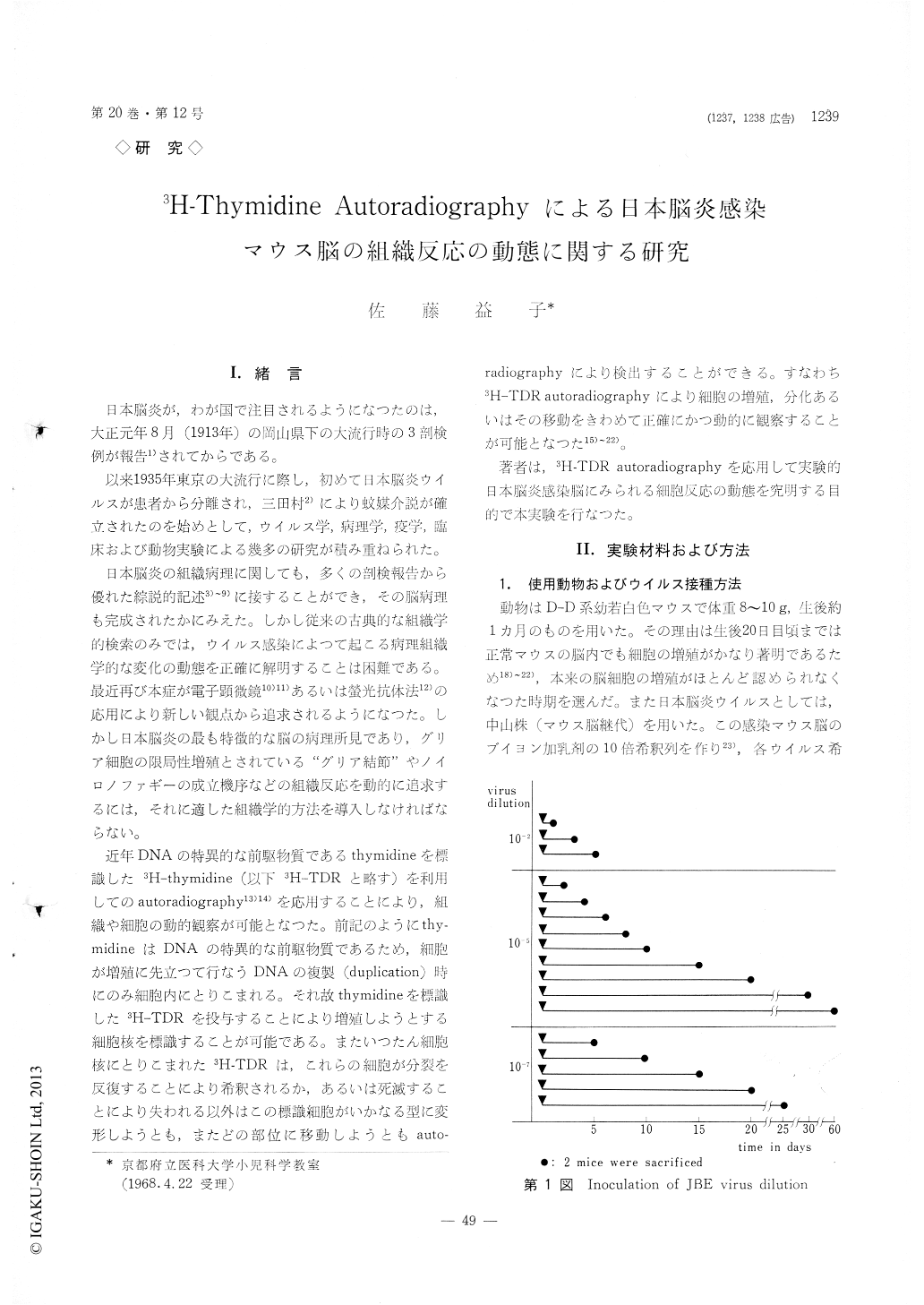
日本脳炎が,わが国で注目されるようになつたのは,大正元年8月(1913年)の岡山県下の大流行時の3剖検例が報告1)されてからである。
以来1935年東京の大流行に際し,初めて日本脳炎ウイルスが患者から分離され,三田村2)により蚊媒介説が確立されたのを始めとして,ウイルス学,病理学,疫学,臨床および動物実験による幾多の研究が積み重ねられた。
Using 3H-thymidine autoradiography, the author investigated origin and fate of reactive cells in the encephalitic lesions of the brain particularly in perivas-cular infiltrations and cellular nodules characteristic of Japanese B encephalitis. First, normal 30-day-old mice were repeatedly injected with 3H-thymidine to confirm that there are few labeled cells in the normal brain except for those in the subependymal layer of the cerebrum. Secondly, mice of the same age were incculated with Japanese B encephalitis virus, and when encephalitis developed, they received a single injection of 3H-thymidine and sacrificed 1 hour later. A few labeled cells were seen in the encephalitic lesion of the brain, but they are not sufficient in number to account for local proliferation of num-erous reactive cells. It was concluded that the reac-tive cells in the brain should he derived from outside of the brain. Thirdly, normal mice of the same age were injected with 3H-thymidine at intervals of 6 hours for 55 hours to label most of circulating leu-cocytes. Two hours after the final injection of 3H-thymidine they were inoculated with Japanese B encephalitis virus and sacrificed at selected intervals. Most cells of the perivascular infiltrations and cel-lular nodules in the encephalitic brain were labeled.
A good correlation was found between labeled per-centage of the reactive cells in the brain and that of circulating leucocytes.
The results of the experiments indicate that leu-cocytes in the peripheral blood, probably mononu-clears, contributed to the formation of perivascular infiltrations and cellular nodules, and that a few leu-cocytes continue to divide locally. Blood mononu-clears seem to be transformed into the so-called "microglia" in the lesions of the brain.
Copyright © 1968, Igaku-Shoin Ltd. All rights reserved.