Japanese
English
- 有料閲覧
- Abstract 文献概要
- 1ページ目 Look Inside
I.緒言
脳腫瘍がいつ頃発生してどのような速さで増殖生長してきたのかということは,われわれが脳腫瘍患者を診るさいに常に関心のもたれるところである。また生長速度あるいは腫瘍細胞の世代時間を知ることは,その腫瘍の過去のヒストリーを明らかにするのみでなく,現時点での増殖生長を未来へ外挿することにより腫瘍の予後を推定する手がかりをあたえる。さらには悪性腫瘍の化学療法を行なうさいに薬剤の投与期間や投与方法を決定するのにおおいに参考になるのである。
腫瘍細胞の生長速度や世代時間を知る方法としては,A)一定時間の前後における細胞数の変化を直接および間接に計測して,増殖曲線を描く,B)腫瘍の体積を直接計測して,大きさの変化を知る,C)分裂指数と分裂時間を計り,細胞増殖の計算式より求める,などの方法があるが3),3H-Thymldineの腫瘍細胞内摂取からその世代時間を算定するのが現在最も信頼できる方法であると思われる。そこでわれわれは3H-Thymidineのin vivo local labelingにより腫瘍細胞のlabeling index(標識率)を求め,これからこの細胞の世代時間を推定し,さらにこれをもとにして2例の幼児にみられた脳腫瘍の発生時期を推測した。
For analysis of the kinetics of cellular proliferation of human brain tumors, 3H-Thymidine has been used in vivo by local application.
METHOD: Tritiated thymidine was injected into the tumor at craniotomy in 0.3ml of 5% glucose solution at a concentration of 100μc per ml. The specimens were obtained about 30 min. after the injection of tritated thymidine reserving blood circu-lation and used for autoradiographic and electron-microscopic studies.
Autoradiographs were prepared by dipping into SAKURA-NR-M2 Emulsion, exposed for 30 days, processed and stained with hematoxylin and eosin.
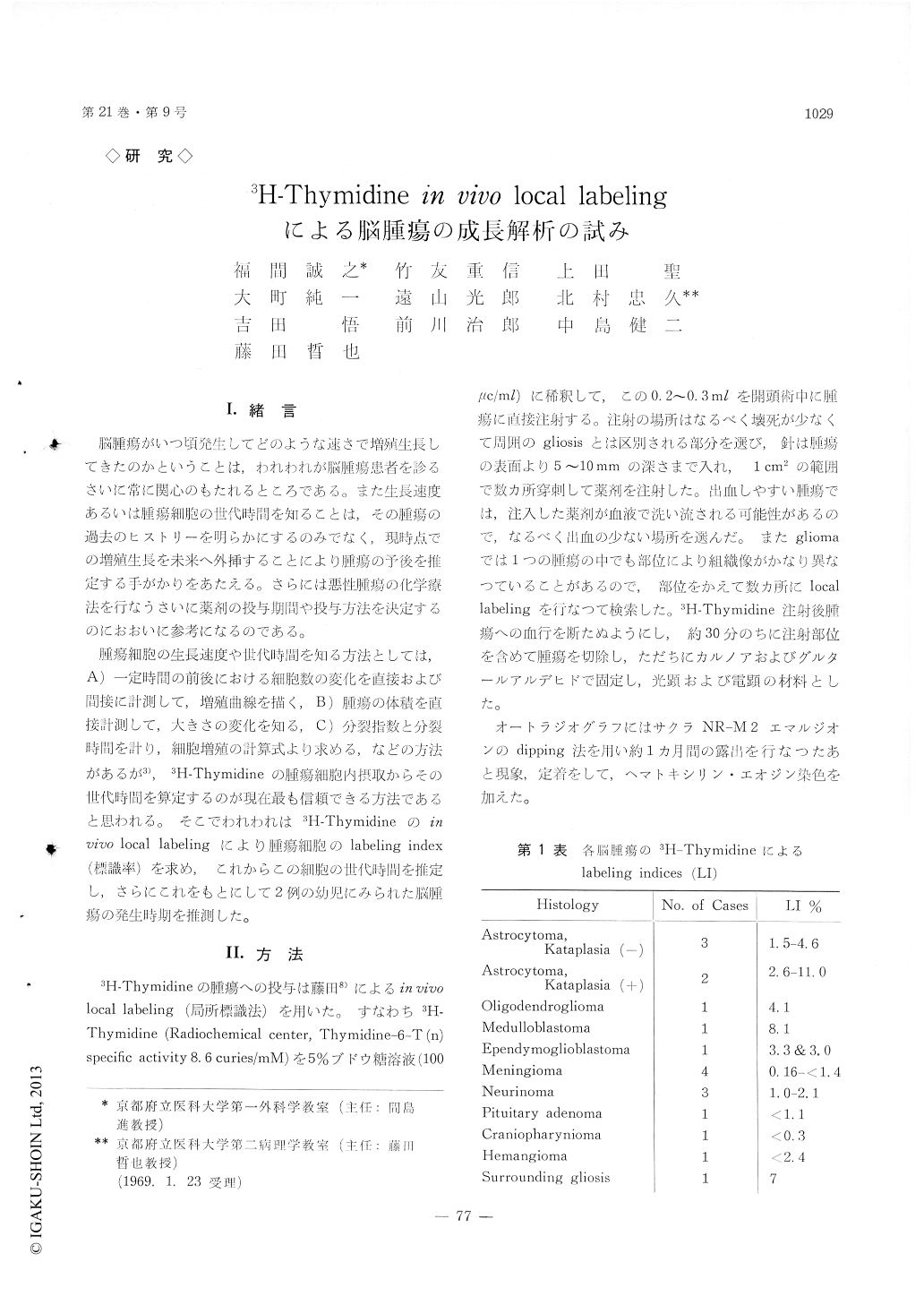
RESULTS: The percentage of labeled cells (Label-ing Index or LI) was counted in various human brain tumors 30 min. after injection on tritiated thymidine. Labeling indices of kataplastic astrocyto-ma were 2.6 to 11.0%. In this type of tumors many endothelial cells showed labeling, but giant cells were not labeled. On the other hand astrocytomas without kataplasia showed LI of 1.6 to 4.6%. The percent-age of labeled tumor cells seems to correlate with the malignancy of the tumors. The labeling index was 4.1% in an oligodendroglioma with astrocyto-matous foci. The foci of astrocytoma in this tumor showed 2.9% of labeling. In meningioma group, LI of the syncytial type was 0.16%, whereas fibro-blastic and angioblastic types showed 1.4% and 0.6%-1.1% respectively. In other brain tumors labeling indices were low ; acoustic neurinomas (1.0-2.1%), pituitary adenoma (under 1.1%) and cranio-pharyngioma (under 0.3%). Higher labeling indices were found for medulloblastoma (8.1%) and open-dymogliohlastoma (3.3%).
By use of labeling index of tumor cells, we tried to analyse the kinetics of cellular proliferation and to infer the time of the onset of the tumors.
Copyright © 1969, Igaku-Shoin Ltd. All rights reserved.