Japanese
English
- 有料閲覧
PPV購入案内
1,320円 (1,200円+税10%) こちらは、353ページから356ページの文献です。購入には医書.jp本会員登録が必要です。
会員登録完了後に改めて上記「購入サイトへ」を選択してください。
または「購入サイトへ」選択後に「購入ログイン」、「新規会員登録」の順に進んでください。
会員登録について詳しくはをご確認ください。
- Abstract 文献概要
- 1ページ目 Look Inside
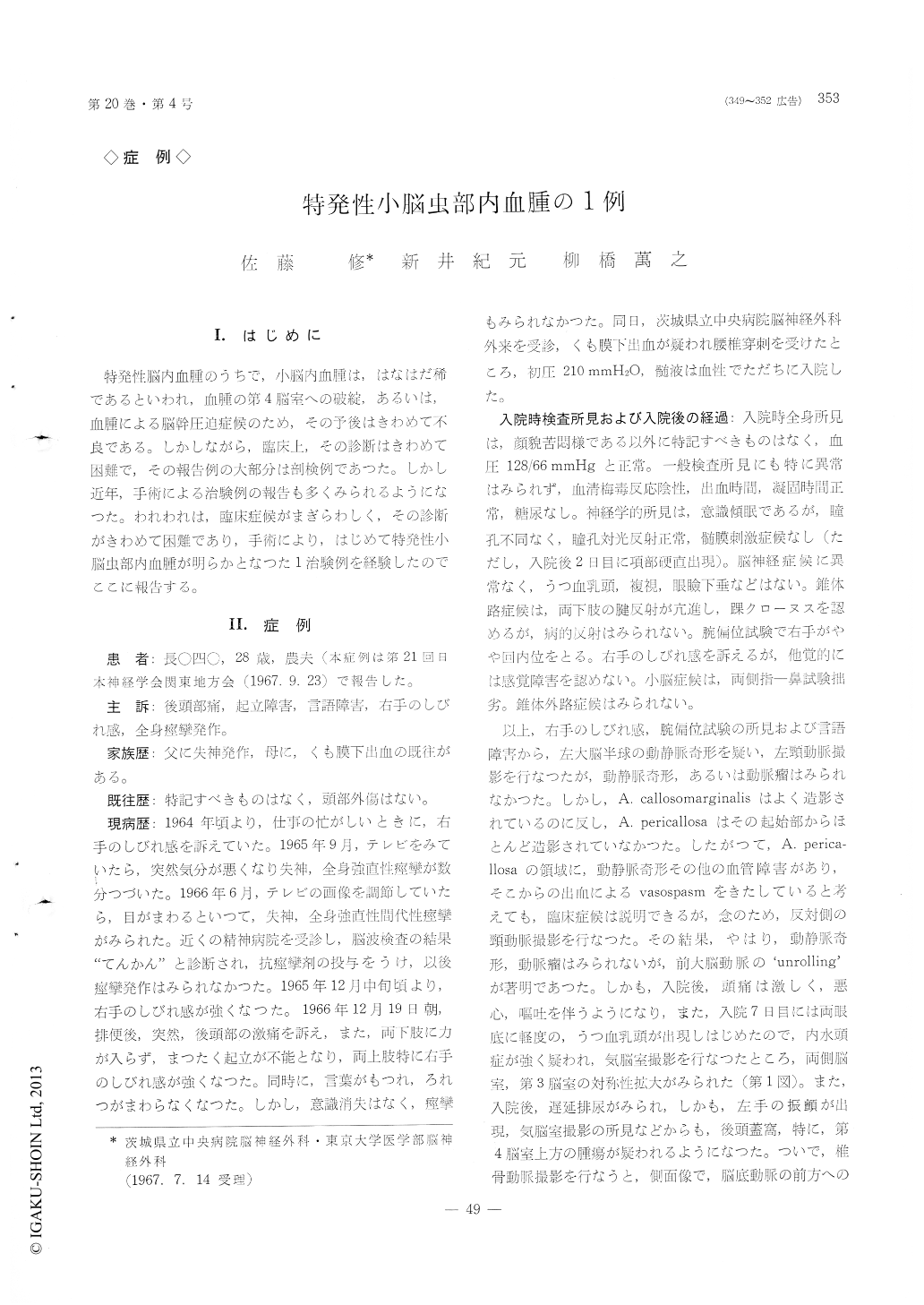
28歳の男子てんかん患者で,突然,後頭部痛,起立障害,言語障害,右手のしびれ感を訴え,腰椎穿刺でくも膜下出血が確認された例を報告した。臨床症候は,左大脳半球の動静脈は奇形が疑われたが,経過を追うにつれ,後頭蓋窩症候が明らかとなり,Myodil脳室撮影で,第4脳室上方の腫瘍が疑われた。後頭下開頭術により,はじめて,小脳虫部内に限局する血腫が発見され,血腫を除去,術後の経過は順調であつた。この例の病因は,おそらく,血管腫の破綻による血腫と考えられた。
一般に,特発性小脳内血腫の臨床的診断は困難と思われる。
The authors reported a young healthy man with spells of epileptic seuzure, who suddenly developed severe occipital headache with akinesia of legs, dys-esthesia of the right arm and speech disturbance. After the lumbar subarachnoid puncture the subarachnoid hemorrhage was ascertained.
In the beginning, the clinical symptoms and signs were confusable with the supratentorial lesion, but in due course of the time the space occupying lesion in the posterior fossa was suggestive. Myodil ventriculogram demonstrated the deformity of the fourth ventricle suggesting the presence of the tumor above the ventricle.
At suboccipital craniectomy, a large cavity con-taining the clot was discovered in the cerebellar vermis. The clot was removed and the postoperative recovery was uneventful.
The cause of the hematoma was considered as the angiomatous malformation, though it was not dis-covered by microscopic examination of the piece of the wall of the cavity.
The authors reviewed the literatures and stressed upon the difficulty of the correct diagnosis of the spontaneous cerebellar hematoma before autopsy or operation.
Copyright © 1968, Igaku-Shoin Ltd. All rights reserved.