Japanese
English
- 有料閲覧
- Abstract 文献概要
- 1ページ目 Look Inside
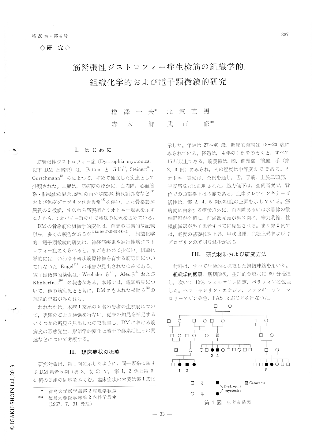
I.はじめに
筋緊張性ジストロフィー症(Dystrophia myotonica,以下DMと略記)は,BattenとGibb5),Steinert39),Curschmann8)らによつて,初めて独立した疾患として分類された。本症は,筋病変のほかに,白内障,心血管系・肺機能の異常,諸種の内分泌障害,糖代謝異常など35)および免疫グロブリン代謝異常48)を伴い,また骨格筋が異質の2徴候,すなわち筋萎縮とミオトニー現象を示すことから,ミオパチー群の中で特殊の位置を占めている。
DMの骨格筋の組繊学的変化は,前記の古典的な記載以来,多くの報告があるが1)12)16)17)29)31)38)49),組織化学的,電子顕微鏡的研究は,神経筋疾患や進行性筋ジストロフィー症にくらべると,まだきわめて少ない。組識化学的には,いわゆる輪状筋原線維を行する筋線維について行なつたEngel11)の報告が見出されたのみである。電子顕微鏡的検索は,Wechslerら46),Aleuら3)およびKlinkerfuss20)の報告がある。本邦では,電顕所見について,他の筋疾患とともに,DMにもふれた松岡ら25)の綜説的記載がみられる。
Histological, histochemical and electron micro-scopic examinations were performed on the thener muscle obtained by biopsy from five patients with myotonic dystrophy from the same pedigree of fami-ly.
Increase of the muscular nuclei with frequent formation of nuclear chain and aggregate especially in the central parts of fibers was a prominent fea-ture by light microscope. However, no fine struc-tural alteration of the nucleus was found except ir-regular and deep imaginations of the nuclear mem-brane. These findings were considered to be a result from amitotic nuclear division and no to be a de-generative change.
Various stages of myofibrillar atrophy were ob-served in almost all muscle fibers affected, and ap-peared to be principal process of the muscular change, at least morphologically. This change se-emed to be caused by gradual destruction of myo-filaments which would commence generally at I-band and peripheral portion of sarcomere. Complete disappearance of the myofibrils extending over several sarcomeres were also found. Nevertheless, the over all arrangement of sarcomere as a functional unit was hardly distorted until late stage of the wasting. The atrophy of the contractile element was some-times more advanced at the subsarcolemmal and perinuclear portions, where scattered fragments of Z substance and myofilament were seen with occa-sional evidence of sarcoplasmic swelling. The second change of myofibrils was a homogenization with obliteration of filamentous structure. The homoge-nization, which appeared to correspond to the light microscopic hyalinization change, showed frequent involvement of the adjoining mitochondria, sarcoplas-mic reticula and sarcoplasms.
Mitochondria showed swelling or shrinkage and sarcoplasmic reticula were dilated sometimes forming large vacuoles. The degeneration of the mitochon-drion and sarcoplasmic reticulum was less marked as compared with myofibrillar changes, and mostly confined to the areas of advanced myofibrillar atrophy or hyalinization. Along these findings, the activities of succinic, lactic dehydrogenases and glutamic ox-aloacetic transaminase were well preserved his-tochemically in the moderately atrophic muscle fibers. However, muscle fibers that were highly atrophic or partly hyalinized showed marked decrease in the activity of these enzymes.
There was no singificant alteration in the fine structure of myoneural junctions. Nonspecific es-terase activity was demonstrated at the location cor-responding to this structure in the majority of the affected muscles. In one patient, proliferation of the fibrous capsule of the muscle spindle was demon-strated.
It was difficult to explain myotonic phenomenon of the disease based on the above described mor-phological changes.
Copyright © 1968, Igaku-Shoin Ltd. All rights reserved.