Japanese
English
- 有料閲覧
PPV購入案内
1,320円 (1,200円+税10%) こちらは、823ページから832ページの文献です。購入には医書.jp本会員登録が必要です。
会員登録完了後に改めて上記「購入サイトへ」を選択してください。
または「購入サイトへ」選択後に「購入ログイン」、「新規会員登録」の順に進んでください。
会員登録について詳しくはをご確認ください。
- Abstract 文献概要
- 1ページ目 Look Inside
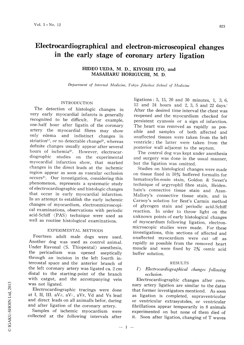
Electrocardiographic and morphological changes in the early myocardial infarction were presented in 14 adult male dogs.
1) On even the earliest stage of myo-cardial ischemia, when hiterto, a detectable change has never been recognized in the histological examination, remarkable pat-hological changes of the myocardium i.e. swelling of mitochondrias, destruction of myofibrills, lightending of sarcoplasma and intacellular edema appear on the electron-microscopic figures.
2) Comparing electrocardiographical al-terations with histological changes in early myocardial infarction, the more intense the myocardial swelling and edema of the affected region progress, the larger the PS-T segment deviation in E.C.G. becomes ; then the severer the coagulation necrosis, the smaller the RS-T segment deviation. The "cove-plane T wave" or the "coronary T wave" in electrocardiogram developes at the end of the coagulation necrosis stadium of morphological examination.
3) It seems that stages of electrocardio-graphic alterations after coronary occlusion correspond to stadiums of morphological changes in the affected myocardium in our experiment : i, e, on the acute stage of electrocardiographic examination, the myocardium shows the figure of the initial stadium of the coagulation necrosis stadium of histological examination ; on the subacute stage of E.C.G., the stadium of cellular infiltration ; on the chronic stage of E.C.G., the stadium for healing, respectively.
Prof. F. Takagi, Director of the Department of Pathology, Tokyo Jikeikai School of Medicine, provided technical assistance on the electronmi-croscopical examination in this experiment.
Copyright © 1957, Igaku-Shoin Ltd. All rights reserved.