Japanese
English
- 有料閲覧
PPV購入案内
1,320円 (1,200円+税10%) こちらは、597ページから610ページの文献です。購入には医書.jp本会員登録が必要です。
会員登録完了後に改めて上記「購入サイトへ」を選択してください。
または「購入サイトへ」選択後に「購入ログイン」、「新規会員登録」の順に進んでください。
会員登録について詳しくはをご確認ください。
- Abstract 文献概要
- 1ページ目 Look Inside
- 参考文献 Reference
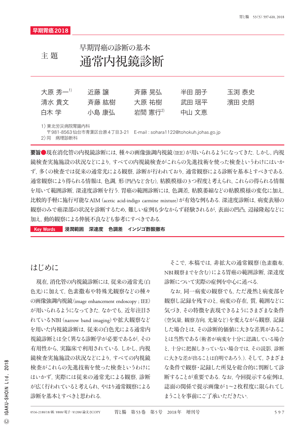
要旨●現在消化管の内視鏡診断には,種々の画像強調内視鏡(IEE)が用いられるようになってきた.しかし,内視鏡検査実施施設の状況などにより,すべての内視鏡検査がこれらの先進技術を使った検査というわけにはいかず,多くの検査では従来の通常光による観察,診断が行われており,通常観察による診断を基本とすべきである.通常観察により得られる情報は,色調,形(凹凸など含む),粘膜模様の3つ程度と考えられ,これらの得られる情報を用いて範囲診断,深達度診断を行う.胃癌の範囲診断には,色調差,粘膜萎縮などの粘膜模様の変化に加え,比較的手軽に施行可能なAIM(acetic acid-indigo carmine mixture)が有効な例もある.深達度診断は,病変表層の観察のみで癌深部の状況を診断するため,難しい症例も少なからず経験されるが,表面の凹凸,辺縁隆起などに加え,動的観察による伸展不良なども参考にすべきである.
A wide range of image-enhanced endoscopy techniques exist for gastrointestinal tract diagnoses. However, not all these advanced technologies are available for endoscopic examinations in specific institutions. Most institutions, in fact, conduct examinations and diagnoses by the conventional method using normal light ; thus, we consider the normal light examination as a standard. Information that can normally be obtained during an endoscopic examination includes the color tone, shape(including unevenness), and mucosal pattern. Diagnosis of the affected area and invasion depth is based on this information. For diagnosis of an area affected by gastric cancer, a relatively simple method involving the use of an acetic acid and indigo carmine mixture is also available depending on the case that allows for the detection of mucosal pattern changes such as color tone differences and mucosal atrophy. However, diagnosis of invasion depth is done by examining only the lesion surface. Therefore, in some cases, diagnosis is difficult, and other variables such as unevenness, marginal ridges, and extension failure, which can all be identified by dynamic observations, should be taken into consideration.
Copyright © 2018, Igaku-Shoin Ltd. All rights reserved.