Japanese
English
- 有料閲覧
PPV購入案内
1,320円 (1,200円+税10%) こちらは、1469ページから1479ページの文献です。購入には医書.jp本会員登録が必要です。
会員登録完了後に改めて上記「購入サイトへ」を選択してください。
または「購入サイトへ」選択後に「購入ログイン」、「新規会員登録」の順に進んでください。
会員登録について詳しくはをご確認ください。
- Abstract 文献概要
- 1ページ目 Look Inside
- 参考文献 Reference
- サイト内被引用 Cited by
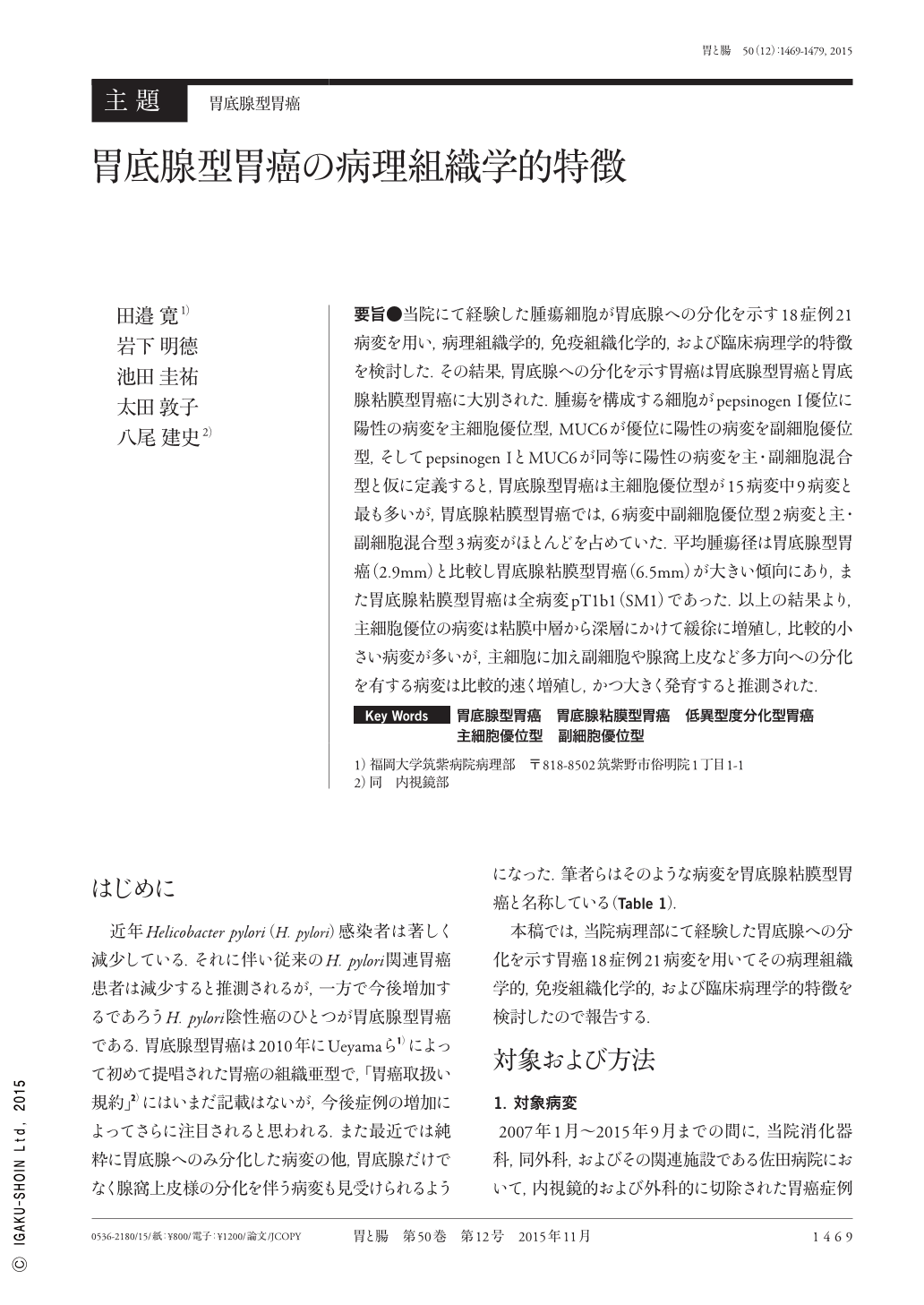
要旨●当院にて経験した腫瘍細胞が胃底腺への分化を示す18症例21病変を用い,病理組織学的,免疫組織化学的,および臨床病理学的特徴を検討した.その結果,胃底腺への分化を示す胃癌は胃底腺型胃癌と胃底腺粘膜型胃癌に大別された.腫瘍を構成する細胞がpepsinogen I優位に陽性の病変を主細胞優位型,MUC6が優位に陽性の病変を副細胞優位型,そしてpepsinogen IとMUC6が同等に陽性の病変を主・副細胞混合型と仮に定義すると,胃底腺型胃癌は主細胞優位型が15病変中9病変と最も多いが,胃底腺粘膜型胃癌では,6病変中副細胞優位型2病変と主・副細胞混合型3病変がほとんどを占めていた.平均腫瘍径は胃底腺型胃癌(2.9mm)と比較し胃底腺粘膜型胃癌(6.5mm)が大きい傾向にあり,また胃底腺粘膜型胃癌は全病変pT1b1(SM1)であった.以上の結果より,主細胞優位の病変は粘膜中層から深層にかけて緩徐に増殖し,比較的小さい病変が多いが,主細胞に加え副細胞や腺窩上皮など多方向への分化を有する病変は比較的速く増殖し,かつ大きく発育すると推測された.
Twenty-one lesions in 18 patients showing differentiation of the tumor cells of the fundic gland observed at the Department of Pathology of the Fukuoka University Chikushi Hospital were studied to evaluate the histopathological, immunohistological, and clinicopathological characteristics of these cells. We found that the gastric adenocarcinomas that differentiate into carcinoma of the fundic gland can be broadly categorized as gastric adenocarcinoma of fundic gland type and gastric adenocarcinoma of fundic mucosa type. When defining lesions comprising tumor cells that are 1)significantly more positive for pepsinogen I, i.e., the chief cell-dominant type, 2)significantly more positive for MUC6, i.e., the mucous neck cell-dominant type, and 3)equally positive for pepsinogen I and MUC6, i.e., the chief-mucous-neck-combination type, we found that those comprising tumor cells of the chief cell-dominant type were the most common, appearing in 9/15 cases of gastric adenocarcinoma of fundic gland type. However, the mucous neck cell-dominant type(2 cases)and chief-mucous-neck- combination type(3 cases)appeared in majority in 6 cases of gastric adenocarcinoma of fundic mucosa type. In terms of average tumor diameters, the tumors of the fundic mucosa type were larger(6.5mm)than those of the fundic gland type(2.9mm). Furthermore, all lesions of the fundic mucosa type represented pT1b1(SM1). These results suggest that lesions of the chief cell-dominant type are relatively small and gradually spread from the middle to the deeper layers of the mucosa, whereas those of the chief cells, mucous neck cells, and foveolar epithelial cells that differentiate in many directions rapidly spread to form large tumors.
Copyright © 2015, Igaku-Shoin Ltd. All rights reserved.