Japanese
English
- 有料閲覧
- Abstract 文献概要
- 1ページ目 Look Inside
- 参考文献 Reference
- サイト内被引用 Cited by
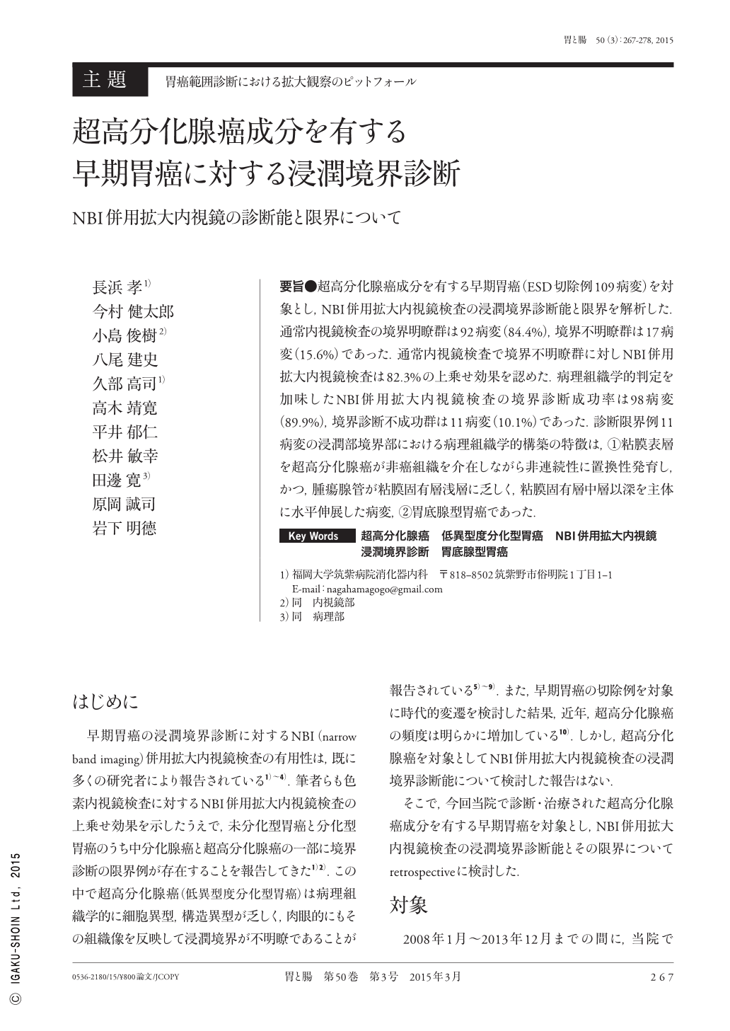
要旨●超高分化腺癌成分を有する早期胃癌(ESD切除例109病変)を対象とし,NBI併用拡大内視鏡検査の浸潤境界診断能と限界を解析した.通常内視鏡検査の境界明瞭群は92病変(84.4%),境界不明瞭群は17病変(15.6%)であった.通常内視鏡検査で境界不明瞭群に対しNBI併用拡大内視鏡検査は82.3%の上乗せ効果を認めた.病理組織学的判定を加味したNBI併用拡大内視鏡検査の境界診断成功率は98病変(89.9%),境界診断不成功群は11病変(10.1%)であった.診断限界例11病変の浸潤部境界部における病理組織学的構築の特徴は,(1)粘膜表層を超高分化腺癌が非癌組織を介在しながら非連続性に置換性発育し,かつ,腫瘍腺管が粘膜固有層浅層に乏しく,粘膜固有層中層以深を主体に水平伸展した病変,(2)胃底腺型胃癌であった.
The invasive margin diagnostic abilities and limitations of magnifying endoscopy with NBI(narrow band imaging)were analyzed in subjects with early-stage gastric cancer(109 lesions in endoscopic submucosal dissection resection specimens)exhibiting adenocarcinoma components with extremely high differentiation. Based on normal endoscopy findings, subjects were divided into a clear margin group(93 lesions; 84.5%)and a poorly defined margin group(17 lesions; 15.5%). Compared with normal endoscopy, NBI magnifying endoscopy achieved additional positive effects in 82.3% of the subjects in the poorly defined margin group. The success rate of margin diagnosis by a combination of NBI magnifying endoscopy and histopathological diagnosis was 89.9%(98 lesions)and the failure rate was 10.1%(11 lesions). In the 11 lesions with a limited diagnosis, the characteristics of the histopathological structures at the invasive margins were(1)adenocarcinoma with extremely high differentiation interspersed with nongastric tissue growing discontinuously in mucosal lamina propria and tumor ducts with horizontal extension through the deeper layers of the mucosal lamina propria, and(2)gastric adenocarcinoma of the fundic gland type.

Copyright © 2015, Igaku-Shoin Ltd. All rights reserved.


