Japanese
English
- 有料閲覧
- Abstract 文献概要
- 1ページ目 Look Inside
- 参考文献 Reference
- サイト内被引用 Cited by
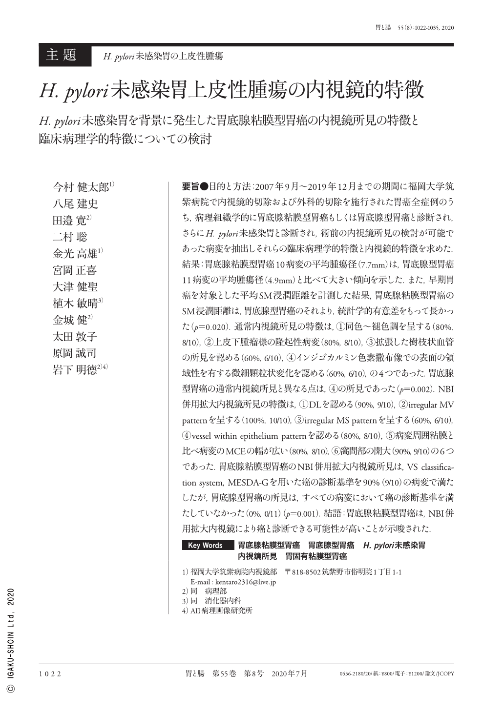
要旨●目的と方法:2007年9月〜2019年12月までの期間に福岡大学筑紫病院で内視鏡的切除および外科的切除を施行された胃癌全症例のうち,病理組織学的に胃底腺粘膜型胃癌もしくは胃底腺型胃癌と診断され,さらにH. pylori未感染胃と診断され,術前の内視鏡所見の検討が可能であった病変を抽出しそれらの臨床病理学的特徴と内視鏡的特徴を求めた.結果:胃底腺粘膜型胃癌10病変の平均腫瘍径(7.7mm)は,胃底腺型胃癌11病変の平均腫瘍径(4.9mm)と比べて大きい傾向を示した.また,早期胃癌を対象とした平均SM浸潤距離を計測した結果,胃底腺粘膜型胃癌のSM浸潤距離は,胃底腺型胃癌のそれより,統計学的有意差をもって長かった(p=0.020).通常内視鏡所見の特徴は,①同色〜褪色調を呈する(80%,8/10),②上皮下腫瘤様の隆起性病変(80%,8/10),③拡張した樹枝状血管の所見を認める(60%,6/10),④インジゴカルミン色素撒布像での表面の領域性を有する微細顆粒状変化を認める(60%,6/10),の4つであった.胃底腺型胃癌の通常内視鏡所見と異なる点は,④の所見であった(p=0.002).NBI併用拡大内視鏡所見の特徴は,①DLを認める(90%,9/10),②irregular MV patternを呈する(100%,10/10),③irregular MS patternを呈する(60%,6/10),④vessel within epithelium patternを認める(80%,8/10),⑤病変周囲粘膜と比べ病変のMCEの幅が広い(80%,8/10),⑥窩間部の開大(90%,9/10)の6つであった.胃底腺粘膜型胃癌のNBI併用拡大内視鏡所見は,VS classification system,MESDA-Gを用いた癌の診断基準を90%(9/10)の病変で満たしたが,胃底腺型胃癌の所見は,すべての病変において癌の診断基準を満たしていなかった(0%,0/11)(p=0.001).結語:胃底腺粘膜型胃癌は,NBI併用拡大内視鏡により癌と診断できる可能性が高いことが示唆された.
Aim and Methods:We collected data regarding lesions in all cases of gastric cancer for which endoscopic or surgical resection was performed at Fukuoka University Chikushi Hospital between September 2007 and December 2019, and which were histopathologically diagnosed as either gastric adenocarcinoma of the fundic gland type or gastric adenocarcinoma of the fundic gland mucosa type. Moreover, the cases analyzed were confirmed as being free of Helicobacter pylori infection and endoscopic findings were available. We investigated the clinical pathological characteristics and characteristic endoscopic findings.
Results:The mean tumor diameter in the fundic gland mucosa type(10 lesions, 7.7mm)tended to be larger than that in the fundic gland type(11 lesions, 4.9mm). Upon measuring the mean submucosal infiltration depths in the cases of early gastric cancer, we found that this depth was statistically significantly deeper in the fundic gland mucosa type than in the fundic gland type(p=0.020). There were 4 characteristic findings noted on normal endoscopy:(1)same/pale color(80% ; 8/10),(2)a elevated lesion resembling a subepithelial tumor(80% ; 8/10),(3)visible branching vessels(60% ; 6/10), and(4)well demarcated area with fine granular surface change visible on the surface with chromoscopy(60% ; 6/10). These findings differed from those for the fundic gland type in that well demarcated area with fine granular surface change could be observed on the surface with chromoscopy(p=0.002). Characteristic findings with NBI magnification endoscopy were(1)presence of a demarcation line(90% ; 9/10),(2)an irregular microvascular pattern(100% ; 10/10),(3)an irregular microsurface pattern(60% ; 6/10),(4)a vessel within epithelium pattern(80% ; 8/10),(5)a wider marginal crypt epithelium pattern than in the surrounding membrane(80% ; 8/10), and(6)wider intervening part between gastric crypts(90% ; 9/10). Although the NBI magnification endoscopy findings for the fundic gland mucosa lesions fulfilled the diagnostic criteria for cancer as described in the vessel plus surface classification system and the magnifying endoscopy simple diagnostic algorithm for early gastric cancer and in 90% of the cases, none of the findings from the fundic gland type lesions fulfilled these criteria(0% ; 0/11)(p=0.001).
Conclusion:Our results demonstrated that it is highly likely that gastric adenocarcinoma of fundic gland mucosa type can be diagnosed as cancer by magnifying endoscopy with NBI.

Copyright © 2020, Igaku-Shoin Ltd. All rights reserved.


