Japanese
English
- 有料閲覧
PPV購入案内
1,320円 (1,200円+税10%) こちらは、1527ページから1541ページの文献です。購入には医書.jp本会員登録が必要です。
会員登録完了後に改めて上記「購入サイトへ」を選択してください。
または「購入サイトへ」選択後に「購入ログイン」、「新規会員登録」の順に進んでください。
会員登録について詳しくはをご確認ください。
- Abstract 文献概要
- 1ページ目 Look Inside
- 参考文献 Reference
- サイト内被引用 Cited by
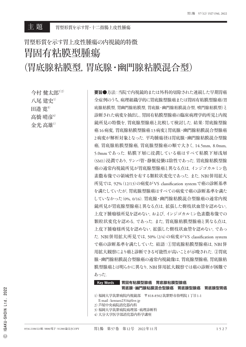
要旨●方法:当院で内視鏡的または外科的切除された連続した早期胃癌全症例のうち,病理組織学的に胃底腺型腺癌または胃固有粘膜型腺癌(胃底腺粘膜型,胃幽門腺粘膜型,胃底腺・幽門腺粘膜混合型,噴門腺粘膜型)と診断された病変を抽出し,胃固有粘膜型腺癌の臨床病理学的所見と内視鏡所見の特徴を,胃底腺型腺癌と比較して検討した.結果:胃底腺型腺癌16病変,胃底腺粘膜型腺癌13病変と胃底腺・幽門腺粘膜混合型腺癌2病変が解析対象となった.平均腫瘍径は胃底腺・幽門腺粘膜混合型腺癌,胃底腺粘膜型腺癌,胃底腺型腺癌の順で大きく,14.5mm,8.0mm,5.0mmであった.粘膜下層に浸潤している癌はすべて粘膜下層浅層(SM1)浸潤であり,リンパ管・静脈侵襲は陰性であった.胃底腺粘膜型腺癌の通常内視鏡所見が胃底腺型腺癌と異なる点は,インジゴカルミン色素撒布像での領域性を有する顆粒状変化であった.また,NBI併用拡大所見では,92%(12/13)の病変がVS classification systemで癌の診断基準を満たしていたが,胃底腺型腺癌はすべての病変で癌の診断基準を満たしていなかった(0%,0/16).胃底腺・幽門腺粘膜混合型腺癌の通常内視鏡所見が胃底腺型腺癌と異なる点は,拡張した樹枝状血管を認めない,上皮下腫瘤様所見を認めない,および,インジゴカルミン色素撒布像での顆粒状変化を認める,であった.また,胃底腺粘膜型腺癌と異なる点は,上皮下腫瘤様所見を認めない,拡張した樹枝状血管を認めない,であった.NBI併用拡大所見では,50%(2/4)の病変がVS classification systemで癌の診断基準を満たしていた.結語:①胃底腺粘膜型腺癌は,NBI併用拡大観察により癌と診断できる可能性が高いことが示唆された.②胃底腺・幽門腺粘膜混合型腺癌の通常内視鏡像は,胃底腺型腺癌,胃底腺粘膜型腺癌とは明らかに異なり,NBI併用拡大観察では癌の診断が困難であった.
This study included patients with early gastric cancer who were treated at Fukuoka University Chikushi Hospital. Patients histologically diagnosed with GA-FG(gastric adenocarcinoma of fundic-gland type), GA-FGM(fundic-gland mucosa type), GA-PGM(pyloric-gland mucosa type), GA-FPM(mixed fundic and pyloric- mucosa type), and GA-CGM(cardiac-gland mucosa type)were enrolled. Histopathological and endoscopic findings were analyzed, including 16 GA-FG, 13 GA-FGM, and 2 GA-FPM lesions. The mean tumor size was larger in GA-FPM, GA-FGM, and GA-FG, at 14.5mm, 8.0mm, and 5.0mm, respectively. Histopathological analysis showed that all lesion types often showed submucosal layer invasion. However, all lesions showed a submucosal invasion distance of <500μm and no lymphatic or venous invasions. Conventional endoscopy using the dye-spraying method revealed the difference between GA-FG and GA-FGM as well-demarcated fine granular areas were observed in GA-FGM lesions. M-NBI(magnifying endoscopy with narrow-band imaging)showed that 12 GA-FGM(92%, 12/13)lesions met the diagnostic criteria for cancer according to the VSCS(vessel plus surface classification system), whereas none of the GA-FG lesions met the same criteria(0%, 0/16). The conventional endoscopic findings of GA-FPM differed from those of GA-FG in the absence of subepithelial tumor-like lesions and dilated branching vessels and the presence of granular changes using the dye-spraying method. The conventional endoscopic findings of GA-FPM differed from those of GA-FGM in the absence of subepithelial tumor-like lesions and dilated branching vessels. M-NBI showed that 2(50%, 2/4)lesions met the VSCS diagnostic criteria for cancer.
Our results suggest that M-NBI is a potentially useful method for GA-FGM diagnosis. The conventional endoscopic findings of GA-FPM were different from those of GA-FG and GA-FGM, and diagnosing cancer by M-NBI was difficult.
Copyright © 2022, Igaku-Shoin Ltd. All rights reserved.