Japanese
English
- 有料閲覧
- Abstract 文献概要
- 1ページ目 Look Inside
- 参考文献 Reference
- サイト内被引用 Cited by
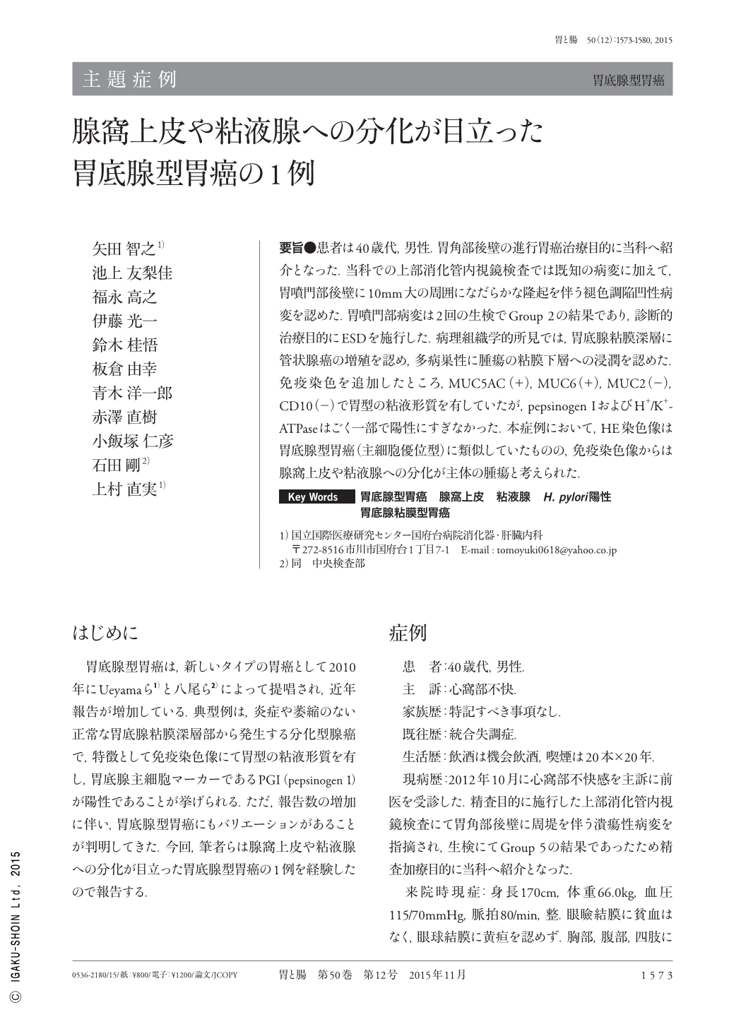
要旨●患者は40歳代,男性.胃角部後壁の進行胃癌治療目的に当科へ紹介となった.当科での上部消化管内視鏡検査では既知の病変に加えて,胃噴門部後壁に10mm大の周囲になだらかな隆起を伴う褪色調陥凹性病変を認めた.胃噴門部病変は2回の生検でGroup 2の結果であり,診断的治療目的にESDを施行した.病理組織学的所見では,胃底腺粘膜深層に管状腺癌の増殖を認め,多病巣性に腫瘍の粘膜下層への浸潤を認めた.免疫染色を追加したところ,MUC5AC(+),MUC6(+),MUC2(−),CD10(−)で胃型の粘液形質を有していたが,pepsinogen IおよびH+/K+- ATPaseはごく一部で陽性にすぎなかった.本症例において,HE染色像は胃底腺型胃癌(主細胞優位型)に類似していたものの,免疫染色像からは腺窩上皮や粘液腺への分化が主体の腫瘍と考えられた.
A male patient in his 40s was referred to our department for treatment of advanced gastric cancer on the posterior wall of the gastric angle. In addition to this known lesion, upper gastrointestinal endoscopy revealed a 10mm, whitish, depressed lesion with a surrounding gentle protrusion on the posterior wall of the gastric cardia. Two biopsy specimens taken from the lesion revealed Group 2(indefinite for neoplasia). The patient then underwent endoscopic submucosal dissection for diagnostic treatment. Histopathological findings indicated proliferation of tubular adenocarcinoma in the deep layers of the mucosa of the fundic gland, in addition to multifocal tumor infiltration into its submucosal layers. Immunostaining revealed that the lesion was MUC5AC(+), MUC6(+), MUC2(−), and CD10(−), indicating a gastric phenotype. However, only very small areas were positive for pepsinogen-I and H+/K+-ATPase. In the present case, although the findings from hematoxylin and eosin staining were similar to those of gastric adenocarcinoma of fundic gland type(chief cell predominant type), immunostaining suggested it to be a tumor related to foveolar epithelium and mucous gland differentiation.

Copyright © 2015, Igaku-Shoin Ltd. All rights reserved.


