Japanese
English
- 有料閲覧
PPV購入案内
1,320円 (1,200円+税10%) こちらは、1362ページから1369ページの文献です。購入には医書.jp本会員登録が必要です。
会員登録完了後に改めて上記「購入サイトへ」を選択してください。
または「購入サイトへ」選択後に「購入ログイン」、「新規会員登録」の順に進んでください。
会員登録について詳しくはをご確認ください。
- Abstract 文献概要
- 1ページ目 Look Inside
- 参考文献 Reference
- サイト内被引用 Cited by
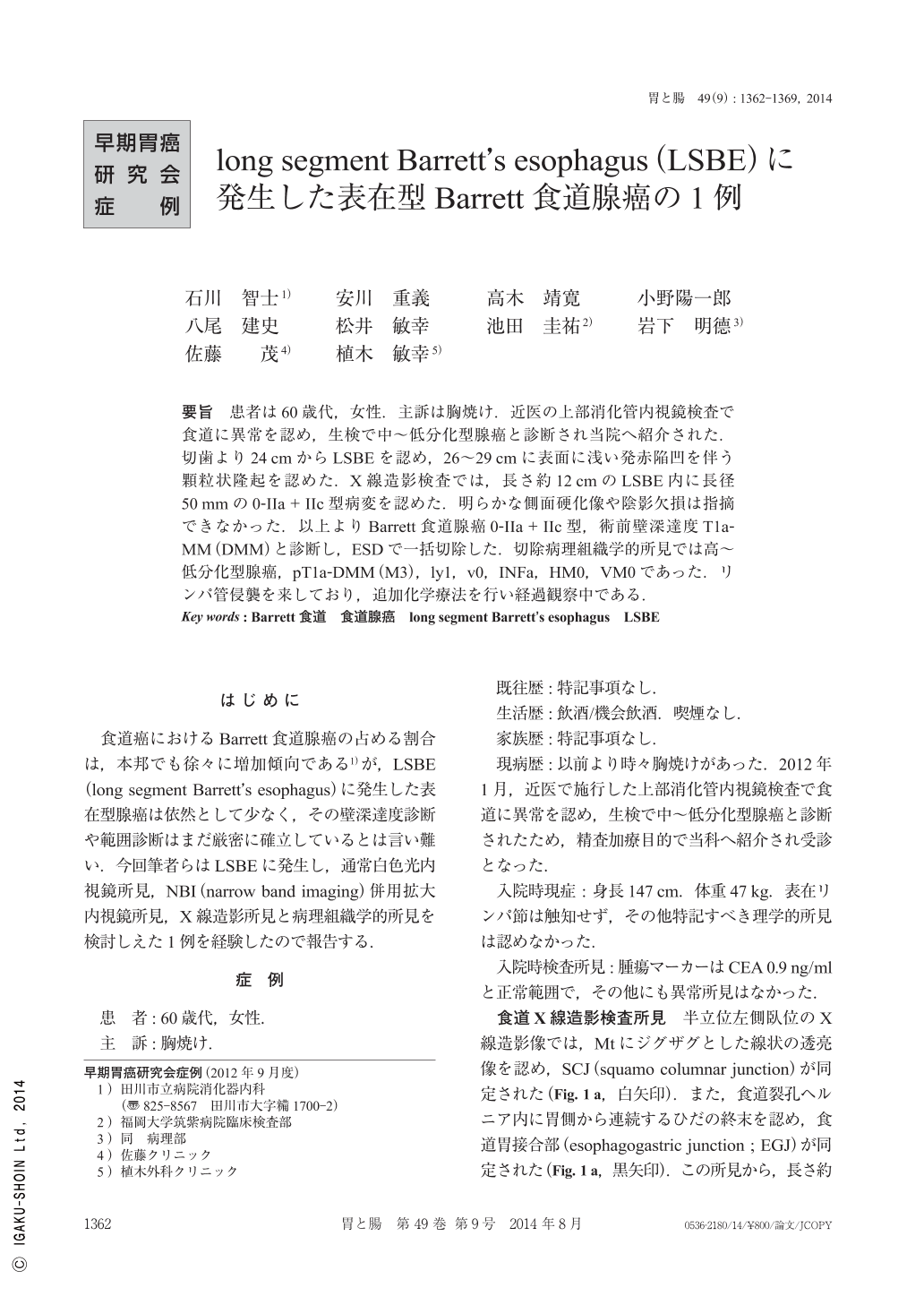
要旨 患者は60歳代,女性.主訴は胸焼け.近医の上部消化管内視鏡検査で食道に異常を認め,生検で中~低分化型腺癌と診断され当院へ紹介された.切歯より24cmからLSBEを認め,26~29cmに表面に浅い発赤陥凹を伴う顆粒状隆起を認めた.X線造影検査では,長さ約12cmのLSBE内に長径50mmの0-IIa+IIc型病変を認めた.明らかな側面硬化像や陰影欠損は指摘できなかった.以上よりBarrett食道腺癌0-IIa+IIc型,術前壁深達度T1a-MM(DMM)と診断し,ESDで一括切除した.切除病理組織学的所見では高~低分化型腺癌,pT1a-DMM(M3),ly1,v0,INFa,HM0,VM0であった.リンパ管侵襲を来しており,追加化学療法を行い経過観察中である.
A 60-year-old woman complained of heartburn. Endoscopy revealed a tumor in the esophagus, and she was referred to our hospital. The tumor was a flat elevated lesion in a long-segment Barrett's esophagus located 26~29cm from the mouth along the total esophageal length. The center of the lesion had a reddish excavation, and an extended hypovascular area was observed around the lesion. The histological findings of a biopsy were consistent with Barrett's esophageal adenocarcinoma. We determined that the tumor invasion depth was within DMM, and performed ESD after obtaining informed consent. The histological findings of the resected specimen were consistent with well to poorly differentiated adenocarcinoma restricted to the mucosa. The tumor invasion depth was confirmed to be within DMM, and lymphatic vessel invasion was also recognized. The patient was treated with chemotherapy and followed up in our hospital.
Copyright © 2014, Igaku-Shoin Ltd. All rights reserved.