Japanese
English
- 有料閲覧
- Abstract 文献概要
- 1ページ目 Look Inside
- 参考文献 Reference
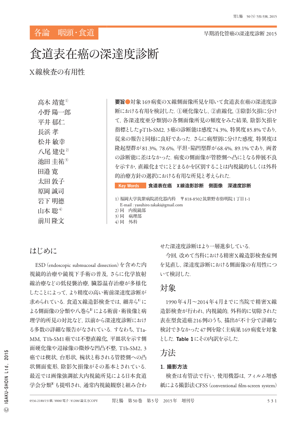
要旨●対象169病変のX線側面像所見を用いて食道表在癌の深達度診断における有用を検討した.(1)硬化像なし,(2)直線化,(3)陰影欠損に分けて,各深達度亜分類別の各側面像所見の頻度をみた結果,陰影欠損を指標としたpT1b-SM2,3癌の診断能は感度74.3%,特異度85.8%であり,従来の報告と同様に良好であった.さらに病型別に分けた感度,特異度は隆起型群が81.3%,78.6%,平坦・陥凹型群が68.4%,89.1%であり,両者の診断能に差はなかった.病変の側面像が管腔側へ凸にとなる伸展不良を示すか,直線化までにとどまるかを区別することは内視鏡的もしくは外科的治療方針の選択における有用な所見と考えられた.
Lateral radiograph findings from 169 lesions were used to investigate the usefulness of these findings in diagnosing the depth of invasion in superficial esophageal cancer. These findings were divided into(1)non-lateral deformity,(2)linearization, and(3)marginal deformity. Results were viewed according to the frequency of each lateral radiograph finding per depth subclassification. We found that the diagnostic capability of pT1b-SM2 and 3 as a marker, when marginal deformity, offered sensitivity of 74.3% and specificity of 85.8%, indicating good results that were consistent with past reports. Furthermore, when divided according to the tumor type, specificity and sensitivity were 81.2% and 78.6%, respectively, for protruding lesions and 68.4% and 89.1%, respectively, for flat and depressed lesions, indicating no difference in the diagnostic capability. The results suggested that using lateral images of lesions to determine whether the lesion exhibiting poor extension protruded into the luminal side or stopped at linearization was useful in selecting the treatment modality, i.e, whether to take an endoscopic or a surgical approach.

Copyright © 2015, Igaku-Shoin Ltd. All rights reserved.


