Japanese
English
- 有料閲覧
- Abstract 文献概要
- 1ページ目 Look Inside
- サイト内被引用 Cited by
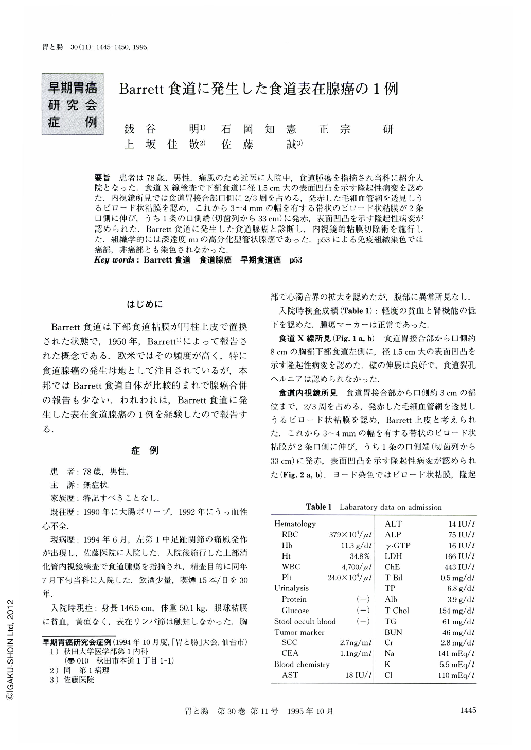
要旨 患者は78歳,男性.痛風のため近医に入院中,食道腫瘍を指摘され当科に紹介入院となった.食道X線検査で下部食道に径1.5cm大の表面凹凸を示す隆起性病変を認めた.内視鏡所見では食道胃接合部口側に2/3周を占める,発赤した毛細血管網を透見しうるビロード状粘膜を認め,これから3~4mmの幅を有する帯状のビロード状粘膜が2条口側に伸び,うち1条の口側端(切歯列から33cm)に発赤,表面凹凸を示す隆起性病変が認められた.Barrett食道に発生した食道腺癌と診断し,内視鏡的粘膜切除術を施行した.組織学的には深達度m3の高分化型管状腺癌であった.p53による免疫組織染色では癌部,非癌部とも染色されなかった.
A 78-year-old male was referred to our department for more detailed evaluation of an esophageal tumor. Upper gastrointestinal x-ray examination revealed an elevated lesion with a granular surface in the lower part of the esophagus. Endoscopic examination revealed smooth-surfaced erythematous mucosa at the oral side of the esophagogastric junction, which connected to a protruding lesion with an irregular surface. A diagnosis of superficial adenocarcinoma arising from Barrett's esophagus led to endoscopic mucosal resection. Histological examination of the resected specimen demonstrated a well differentiated adenocarcinoma arising from columnar epithelium of the esophagus (Barrett's esophagus) and the invasion was limited within the muscularis mucosa (m3). There was no p53 immunoreactivity which was frequently observed in the adenocarcinoma arising from Barrett's esophagus. In addition, 29 cases of early esophageal adenocarcinoma arising from Barrett's esophagus reported in Japan were briefly reviewed.

Copyright © 1995, Igaku-Shoin Ltd. All rights reserved.


