Japanese
English
- 有料閲覧
PPV購入案内
1,320円 (1,200円+税10%) こちらは、915ページから920ページの文献です。購入には医書.jp本会員登録が必要です。
会員登録完了後に改めて上記「購入サイトへ」を選択してください。
または「購入サイトへ」選択後に「購入ログイン」、「新規会員登録」の順に進んでください。
会員登録について詳しくはをご確認ください。
- Abstract 文献概要
- 1ページ目 Look Inside
- 参考文献 Reference
- サイト内被引用 Cited by
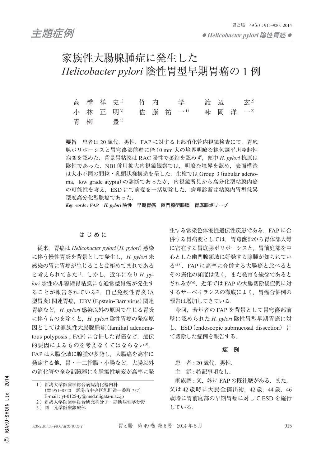
要旨 患者は20歳代,男性.FAPに対する上部消化管内視鏡検査にて,胃底腺ポリポーシスと胃穹窿部前壁に径10mm大の境界明瞭な褪色調平坦隆起性病変を認めた.背景胃粘膜はRAC陽性で萎縮を認めず,便中H. pylori抗原は陰性であった.NBI併用拡大内視鏡観察では,明瞭な境界を認め,表面構造は大小不同の顆粒・乳頭状様構造を呈した.生検ではGroup 3(tubular adenoma,low-grade atypia)の診断であったが,内視鏡所見から高分化型粘膜内癌の可能性を考え,ESDにて病変を一括切除した.病理診断は粘膜内胃型低異型度高分化型腺癌であった.
The patient was a male in his twenties with FAP(familial adenomatous polyposis). Conventional endoscopy revealed fundic gland polyposis spreading over the stomach, except in the antrum, and a white, slightly elevated lesion, approximately 10mm in diameter on the anterior wall of gastric fornix. The background mucosa showed regular arrangement of collecting venules and no atrophy, and stool H. pylori antigen was negative. The surface of the lesion revealed rough and granular patterns of various shapes. Magnifying endoscopy with NBI(narrow band imaging)showed irregular granular/papillary surface structures with loop-shaped microvascular vessels. Though the histopathogical diagnosis of the biopsy specimen was Group 3(tubular adenoma, low-grade atypia), NBI findings were considered to be intramural, well-differentiated, early gastric carcinoma. After obtaining informed consent from the patient, ESD(endoscopic submucosal dissection)was performed. Histologically, the resected specimen was diagnosed as a well-differentiated adenocarcinoma with low grade atypia, which showed a gastric mucin phenotype.
For patients with FAP without H. pylori infection, it is important to investigate the stomach using endoscopy, particularly the gastric body to detect early cancer, as reported in this case.
Copyright © 2014, Igaku-Shoin Ltd. All rights reserved.