Japanese
English
- 有料閲覧
PPV購入案内
1,320円 (1,200円+税10%) こちらは、362ページから368ページの文献です。購入には医書.jp本会員登録が必要です。
会員登録完了後に改めて上記「購入サイトへ」を選択してください。
または「購入サイトへ」選択後に「購入ログイン」、「新規会員登録」の順に進んでください。
会員登録について詳しくはをご確認ください。
- Abstract 文献概要
- 1ページ目 Look Inside
- 参考文献 Reference
- サイト内被引用 Cited by
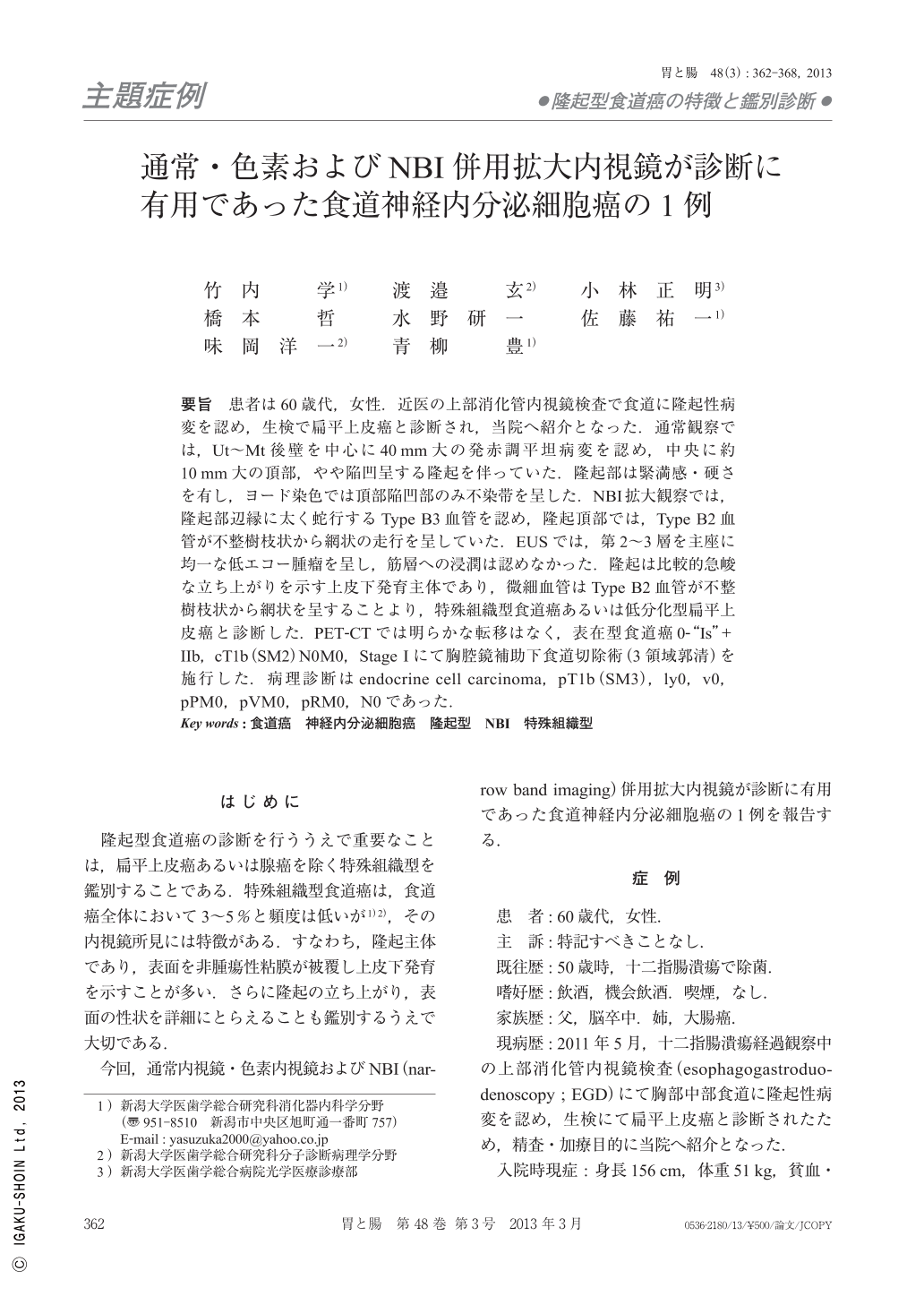
要旨 患者は60歳代,女性.近医の上部消化管内視鏡検査で食道に隆起性病変を認め,生検で扁平上皮癌と診断され,当院へ紹介となった.通常観察では,Ut~Mt後壁を中心に40mm大の発赤調平坦病変を認め,中央に約10mm大の頂部,やや陥凹呈する隆起を伴っていた.隆起部は緊満感・硬さを有し,ヨード染色では頂部陥凹部のみ不染帯を呈した.NBI拡大観察では,隆起部辺縁に太く蛇行するType B3血管を認め,隆起頂部では,Type B2血管が不整樹枝状から網状の走行を呈していた.EUSでは,第2~3層を主座に均一な低エコー腫瘤を呈し,筋層への浸潤は認めなかった.隆起は比較的急峻な立ち上がりを示す上皮下発育主体であり,微細血管はType B2血管が不整樹枝状から網状を呈することより,特殊組織型食道癌あるいは低分化型扁平上皮癌と診断した.PET-CTでは明らかな転移はなく,表在型食道癌0-“Is”+IIb,cT1b(SM2)N0M0,Stage Iにて胸腔鏡補助下食道切除術(3領域郭清)を施行した.病理診断はendocrine cell carcinoma,pT1b(SM3),ly0,v0,pPM0,pVM0,pRM0,N0であった.
A female in her sixties. The conventional esophagoscopy showed a reddish flat elevated lesion and a protruded lesion with central slight depression on the posterior wall of the upper to lower thoracic esophagus. The protruded lesion had overall thickness and hardness. Only the central depression of the protrusion was unstained with iodine. The magnifying endoscopy with NBI revealed Type B2 showing an irregular branched, reticular microvessel(type 4R)without forming AVA and Type B3 consisting of vessels three times or more thicker than Type B2. EUS(IDUS, 20MHz)showed a hypo echoic mass lesion invading to the SM deep layer.
We diagnosed the lesion as a special variant of esophageal carcinoma or poorly differentiated squamous cell carcinoma because the lesion revealed a protrusion covered partly with normal epithelium and irregular branched, reticular vascular pattern. Video-Assisted Thoracic Surgery for the Esophagus with three-field lymph node dissection was performed. Histologically, the tumor was diagnosed as endocrine cell carcinoma pT1b(SM3), ly0, v0, pPM0, pVM0, pRM0, n(-). Immmunohistochemically, CD56 and chromogranin A were positive for the tumor cells.
Copyright © 2013, Igaku-Shoin Ltd. All rights reserved.