Japanese
English
- 有料閲覧
PPV購入案内
1,320円 (1,200円+税10%) こちらは、504ページから508ページの文献です。購入には医書.jp本会員登録が必要です。
会員登録完了後に改めて上記「購入サイトへ」を選択してください。
または「購入サイトへ」選択後に「購入ログイン」、「新規会員登録」の順に進んでください。
会員登録について詳しくはをご確認ください。
- Abstract 文献概要
- 1ページ目 Look Inside
症 例
患 者:M. N. 52歳 女 家婦
主 訴:心窩部不快感
既往歴及び家族歴:特記すべきものなし.
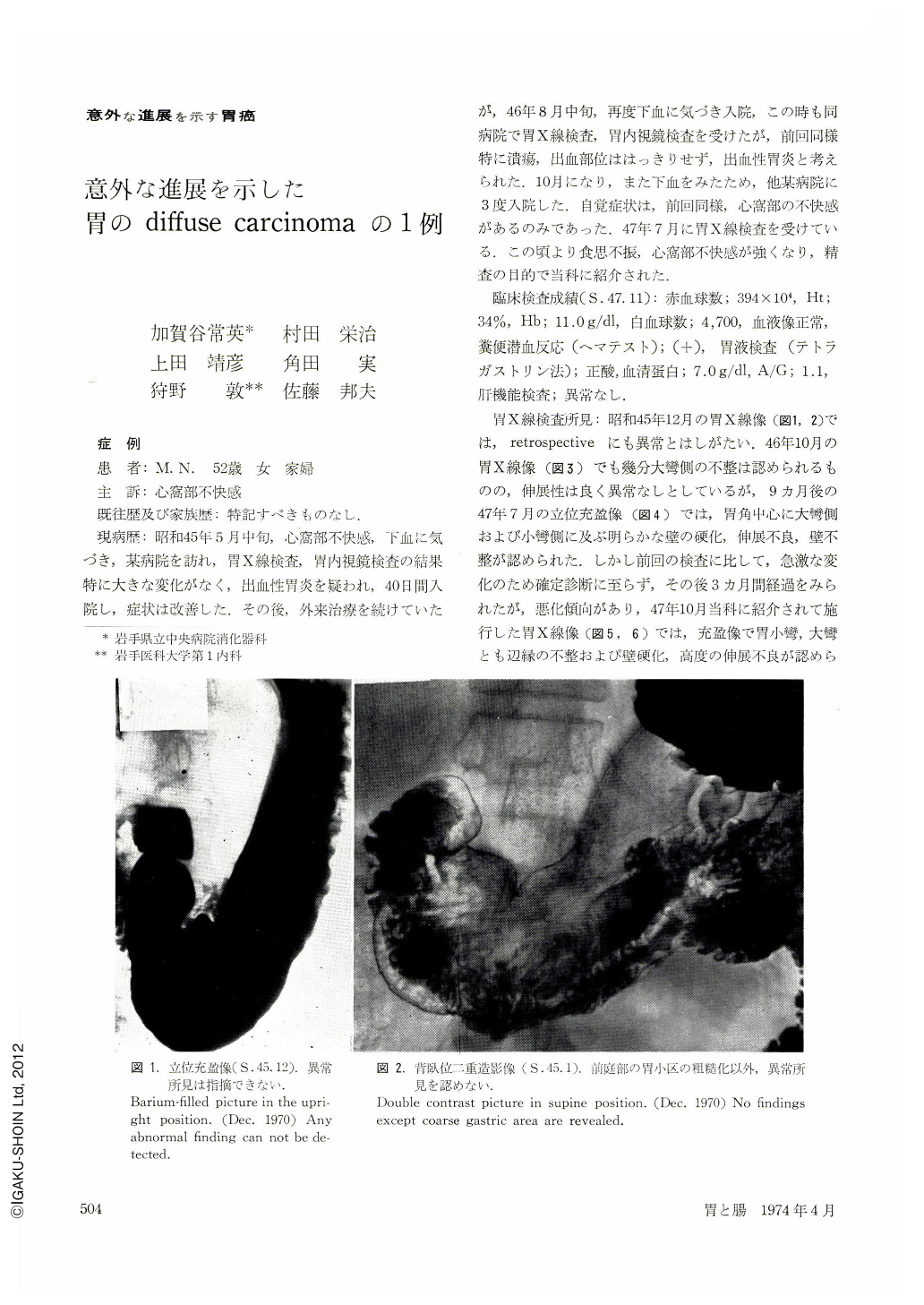
現病歴:昭和45年5月中旬,心窩部不快感,下血に気づき,某病院を訪れ,胃X線検査,胃内視鏡検査の結果特に大きな変化がなく,出血性胃炎を疑われ,40日間入院し,症状は改善した.その後,外来治療を続けていたが,46年8月中旬,再度下血に気づき入院,この時も同病院で胃X線検査,胃内視鏡検査を受けたが,前回同様特に潰瘍,出血部位ははっきりせず,出血性胃炎と考えられた.10月になり,また下血をみたため,他某病院に3度入院した.自覚症状は,前回同様,心窩部の不快感があるのみであった.47年7月に胃X線検査を受けている.この頃より食思不振,心窩部不快感が強くなり,精査の目的で当科に紹介された.
A case of gastric cancer is described here that has been followed up for as long as 29 months since the initial examination, showing a sudden change for the worse within the last nine months.
A 53-year-old woman noticed recurring bouts of unpleasant sensation in the epigastrium along with melena, and she was followed up at a hospital under a diagnosis of hemorrhagic gastritis. In the interim roentgenography and endoscopy of the stomach failed to locate the site of bleeding. She was then treated in another hospital for about one year more. Three months before she was referred to our hospital the stomach showed sudden change in its contour including rigidity, irregularity and loss of distensibility.
Roentgenography prior to surgical intervention revealed marginal irregularity and rigidity of the wall extending to both sides of the curvatures. Lack of distensibility was evident. Endoscopy also showed widened angle, and margins of the mucosal folds of low stature were made out with difficulty. On the greater curvature was noticed a round ulcer with irregularly raised edges. Gastric biopsy demonstrated adenocarcinoma mucocellulare scirrhosum. The patient underwent gastrectomy under a diagnosis of diffuse carcinoma. Findings to operation were S2, P1, H0, N1 according to the General Rules for Gastric Cancer Study in Surgery and Pathology. Cancer cells were mostly found in or beneath the submucosal layer. Only a fraction of Ⅱc was seen in the mucosa around the ulcer; otherwise cancer nests remained unexposed over the surface. Histologically, Ⅱc part was of signet-ring cell type and others, of scirrhous adenocarcinoma variety. Retrospective study of the present case showed that in roentgenography it was not until nine months prior to sudden alterations that a slight change was discernible. On the other hand, the initial pictures of endoscopy showed a minor reddened area at a site corresponding to that of the chief lesion demonstrated in later films. It was assumed that a Ⅱb-like change must have been there already then. It is of great interest were it possible to regard it as the source of diffuse carcinoma.
Copyright © 1974, Igaku-Shoin Ltd. All rights reserved.