Japanese
English
- 有料閲覧
- Abstract 文献概要
- 1ページ目 Look Inside
- サイト内被引用 Cited by
Granular cell myoblastomaは全身のいかなる部位にも発生しうる良性腫瘍で,これまで1,000例近くの報告があり,その好発部位は舌,乳房および全身各所の皮下組織である.消化管における本症の発生は比較的少なく,現在までに食道,胃,十二指腸,大腸,虫垂,直腸,総胆管での発生が報告されている1)~3).しかしこれらは剖検により偶然発見されたか,あるいは術前諸検査により病変の存在のみが指摘され,術後の組織検査ではじめて確定診断の得られたものがほとんどである.
今回われわれは極めて特徴的な内視鏡所見を呈した食道の隆起性病変より生検を施行し,granular cell myoblastomaと診断しえた症例を経験したので,その内視鏡像および本症に関する文献的考察を加えて報告する.
A case of granular cell myoblastoma of the esophagus was reported with review of the world literature. The granular cell myoblastoma is a benign lesion which is founcl anywhere in the body and occured most frequently in the tongue and skin.
To our knowledge only seventeen cases of the tumor were reported in the world literature and the present case was the 18th. Since the granular cell myoblastoma of the esophagus is usually recognized as a submucosal tumor covered with the intact mucosa of the esophagus, accurate preoperative diagnosis is rare. The correct diagnosis of the majority of reported cases were made by the histological examination after surgery.
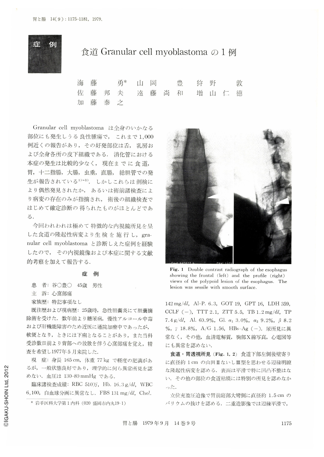
A 45-year-old man was seen at the Iwate Medical University Hospital complaining of mild epigastric pain. An esophagogastrogram demonstrated a polypoid lesion in the distal esophagus and on the antrum of the stomach.
An esophagogastroscopy revealed two white, slightly shiny, well defined elevated lesion on the esophagus. The appearance looked as a molar on the gingiva. Biopsy specimens were taken from the lesions and it was diagnosed as granular cell myoblastoma of the esophagus. Biopsy taken from the gastric lesion revealed normal mucosa only. The ultrastructural findings of the present case were almost same as that of previously published cases. The reason why the present case was diagnosed by routine biopsy method was suspected that the tumor occurred at submucosal layer just beneath the mucosa and grew toward the lumen of the esophagus and resulted the covering mucosa to make thin. The previously reported 17 cases of the tumor and the present case were summerlized in Table 1. The age of the patients ranged from 19 to 59 years and the majority occurred in the patients between 40 to 50 years of age. The tumors occurred frequently at the distal and proximal part of the esophagus. The tumor are occasionally multicentric either in the esophagus or in other part of the body.
Copyright © 1979, Igaku-Shoin Ltd. All rights reserved.