Japanese
English
- 有料閲覧
PPV購入案内
1,320円 (1,200円+税10%) こちらは、437ページから443ページの文献です。購入には医書.jp本会員登録が必要です。
会員登録完了後に改めて上記「購入サイトへ」を選択してください。
または「購入サイトへ」選択後に「購入ログイン」、「新規会員登録」の順に進んでください。
会員登録について詳しくはをご確認ください。
- Abstract 文献概要
- 1ページ目 Look Inside
- サイト内被引用 Cited by
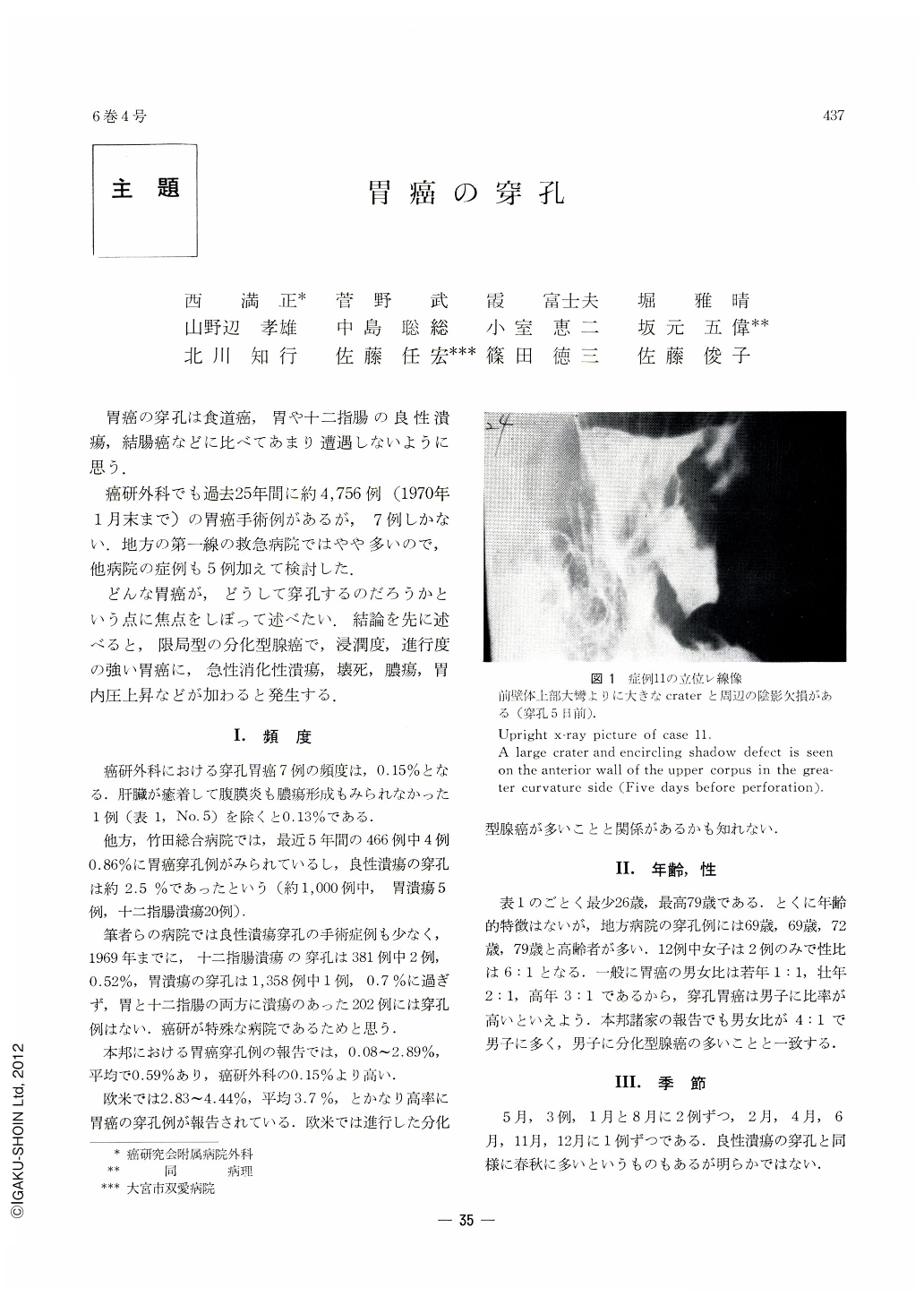
胃癌の穿孔は食道癌,胃や十二指腸の良性潰瘍,結腸癌などに比べてあまり遭遇しないように思う.
癌研外科でも過去25年間に約4,756例(1970年1月末まで)の胃癌手術例があるが,7例しかない.地方の第一線の救急病院ではやや多いので,他病院の症例も5例加えて検討した.
Of 4,576 cases of gastric cancer surgically treated up to the end of January 1970 at the Surgical Department, Cancer Institute Hospital, perforated cases of gastric cancer numbered only 7, or 0.15 per cent. Clinical and pathological review has been tried on these cases together with 5 cases treated in other hospitals.
Gastric perforation was seen far frequently in the male; 10 cases were male and only 2 were female.
Prior to about one week before surgical exploration, the patients used to complain of aggravated pain, vomiting and hematemesis, and many of them had undergone various examinations on these accounts. Surgically the stomach was removed in 7 cases, and closure of perforation in 5. Mostly the patients were in such a weakened state due to advanced carcinoma that prognosis after resection proved very unsatisfactory.
The gastric cancer which caused acute perforation belonged to localized, differentiated medullary adenocarcinoma, showing more rapid degree of infiltration and spread. A cancer nest that tends to be accompanied with necrosis and infection may proliferate in such a manner as to displace or penetrate the muscularis propriae. Such a lesion easily leads to perforation of the gastric wall.
Classified according to Borrmann, perforative cancer mostly belongs to Type Ⅲ. S-, N-, P-, H-factor is also high.
Perforation is caused either by penetration of cancer lesion itself or by that of thin ulcer floor consisting of connective tissues that are free from cancer cells. In either case, no scarification has taken place in the surrounding areas, and the whole picture shows findings of acute ulcer.
Copyright © 1971, Igaku-Shoin Ltd. All rights reserved.