Japanese
English
- 有料閲覧
PPV購入案内
1,320円 (1,200円+税10%) こちらは、1521ページから1525ページの文献です。購入には医書.jp本会員登録が必要です。
会員登録完了後に改めて上記「購入サイトへ」を選択してください。
または「購入サイトへ」選択後に「購入ログイン」、「新規会員登録」の順に進んでください。
会員登録について詳しくはをご確認ください。
- Abstract 文献概要
- 1ページ目 Look Inside
はじめに
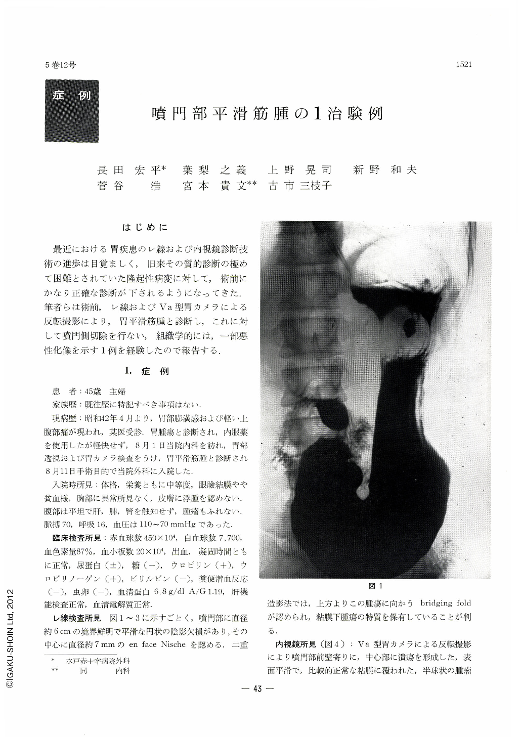
最近における胃疾患のレ線および内視鏡診断技術の進歩は目覚ましく,旧来その質的診断の極めて困難とされていた隆起性病変に対して,術前にかなり正確な診断が下されるようになってきた.筆者らは術前,レ線およびVa型胃カメラによる反転撮影により,胃平滑筋腫と診断し,これに対して噴門側切除を行ない,組織学的には,一部悪性化像を示す1例を経験したので報告する.
By dint of remarkable progress in the fields of x-ray and endoscopy, qualitative diagnosis of protruding lesions in the stomach, hitherto considered to be extremely difiicult, is now made with facility.
This paper illustrates a case of leiomyoma in the cardiac portion preoperatively diagnosed as such associated with partial malignancy histologically proved.
The patient is a 45-year-old woman complaining of feeling of fullness in the epigastric region. At x-ray examination, a smooth, sharply circumscribed shadow defect, measuring about 6cm in diameter, was visualized in the cardiac region. Bridging folds approaching from the oral side toward and over the tumor was also demonstrated by double contrast method. These findings are characteristic of submucosal tumor. Endoscopy revealed at the cardiac region near the anterior wall a hemispheric tumor covered with relatively normal mucosa of smooth surface. Ulceration was seen in its center.
The results of these examinations led the authors to the diagnosis of leiomyoma of the stomach, and accordingly gastric resection on the cardiac side was performed. Postoperative course was uneventful. The patient has been enjoying good health doing her daily chores.
Histologically, typical picture of leiomyoma was seen in the majority of blocks, where muscle fibers were in orderly arrangement. However, in several blocks muscle fibers were disarranged and variation in size of nucleus and its mitosis were recognized. Since as yet pathological criteria of benign and malignant natures of leiomyoma have not been well established, this case is reported as it is as a case of partial malignancy seen in a leiomyoma
Copyright © 1970, Igaku-Shoin Ltd. All rights reserved.