Japanese
English
- 有料閲覧
- Abstract 文献概要
- 1ページ目 Look Inside
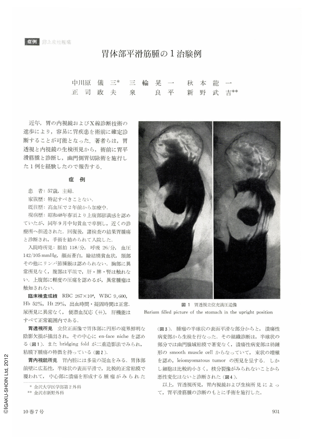
近年,胃の内視鏡およびX線診断技術の進歩により,容易に胃疾患を術前に確定診断することが可能となった.著者らは,胃透視と内視鏡の生検所見から,術前に胃平滑筋腫と診断し,幽門側胃切除術を施行した1例を経験したので報告する.
A report is made of leiomyoma of the gastric body preoperatively diagnosed as such by fluoroscopy and endoscopy of the stomach.
The patient, a housewife 57 years of age, came to the hospital because she had fainted on account of anemia. Fluoroscopy of the stomach showed a well-defined round shadow defect with an en face niche in the center. This finding, together with bridging folds revealed by double contrast picture, was suggestive of a submucosal tumor. Endoscopy also showed a broad-based, hemispheric tumor with central ulcer formation on the anterior wall of the body. Biopsy confirmed the diagnosis of leiomyoma.
Since the majority of gastric leiomyoma develop in the age bracket 40~70, differentiation from cancer is important. Leiomyoma would show multiple occurrence in the body, but it may also develop singly and show a tendency to outward development. In about 5 per cent it is said to become malignant. Accurate diagnosis can be established by biopsy, but, as Nagata et al. has reported, careful attention should be paid to the fact that only in a part of the tumor can malignant change be observed.

Copyright © 1975, Igaku-Shoin Ltd. All rights reserved.


