Japanese
English
- 有料閲覧
- Abstract 文献概要
- 1ページ目 Look Inside
はじめに
十二指腸原発の良性腫瘍としては,腺腫,線維腫,平滑筋腫などがあるが,いずれも比較的まれで,とくに平滑筋腫は胃に発生するものと比較してきわめて珍らしいものとされている.
筆者らは,最近その1例を経験したので,症例を報告するとともに,文献的考察をこころみた.
A case of leiomyoma of the duodenum has been studied.
The patient is a 41-year-old woman who had an episode of right hypochondrial pain and hematemesis in 1960. While working in the field three years later, she was overtaken by dizziness, nausea, epigastric distress and tarry stool. She was admitted to hospital at once. At physical examination, a tumor the size of a duck's egg was palpated in the right upper abdomen.
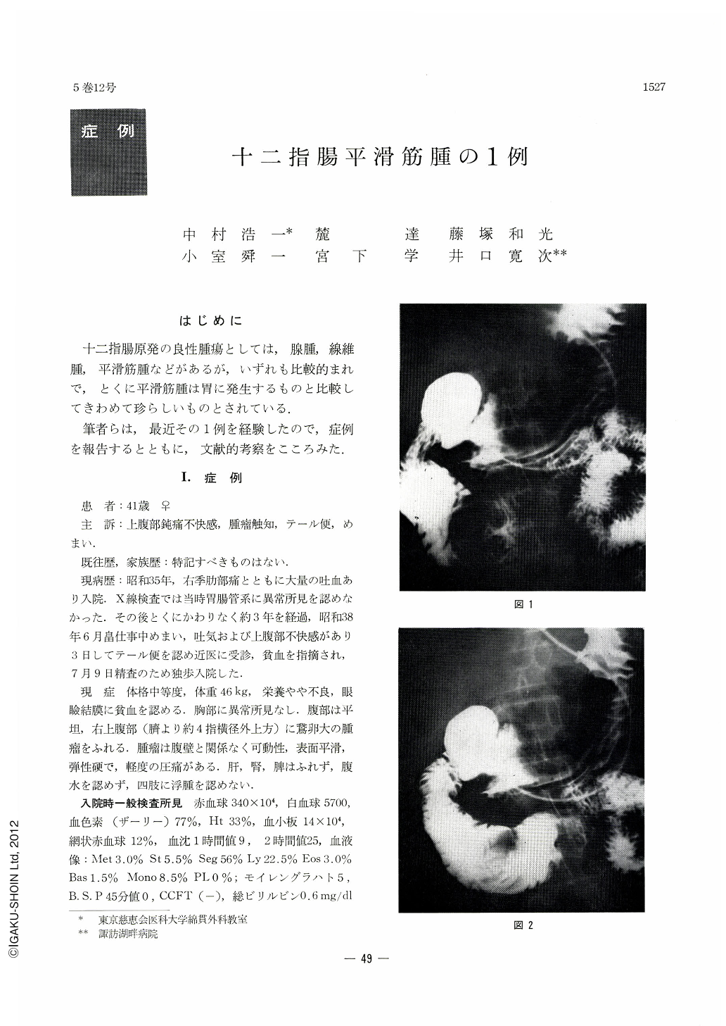
Hypotonic duodenography disclosed a filling shadow defect in the pars horizontalis inferior of the duodenum. After blood transfusion she underwent laparotomy. The posterior wall of the stomach adhered to the pancreas, and one third of the transverse colon distal from the hepatic flexure showed marked adhesion to the pylorus and duodenum. Cleaned of the adhesions, the now mobile tumor was found to be located in the lower horizontal part of the duodenum. When it was opened on the anterior wall, the tumor showed extracanalicular development like a diverticulum. Coagulated blood was seen on the mucosa of the orifice. The base of the tumor showed marked connective-tissue-like adhesion to the retroperitoneal tissues, so that the tumor was cut off from them and excised. Duodenojejunal end-to-end anastomosis was then performed. The removed tumor the size of a mat's knuckle showed a diverticular development having a relatively thick wall comprised of tenacious connective tissues. A great amount of coagulated blood was seen in its inner surface which was covered with granulation tissues. Infiltration into its neighborhood was slight. Histologically, proliferation of smooth muscle fiber bundles was partly seen with round cell infiltration. No nuclear atypicality was observed. The surface of the tumor was involved by necrosis and granulation tissues. In places bleeding was noticed. No infiltration into the duodenal epithelium or other neighboring tissues was recognized.

Copyright © 1970, Igaku-Shoin Ltd. All rights reserved.


