Japanese
English
- 有料閲覧
PPV購入案内
1,320円 (1,200円+税10%) こちらは、293ページから297ページの文献です。購入には医書.jp本会員登録が必要です。
会員登録完了後に改めて上記「購入サイトへ」を選択してください。
または「購入サイトへ」選択後に「購入ログイン」、「新規会員登録」の順に進んでください。
会員登録について詳しくはをご確認ください。
- Abstract 文献概要
- 1ページ目 Look Inside
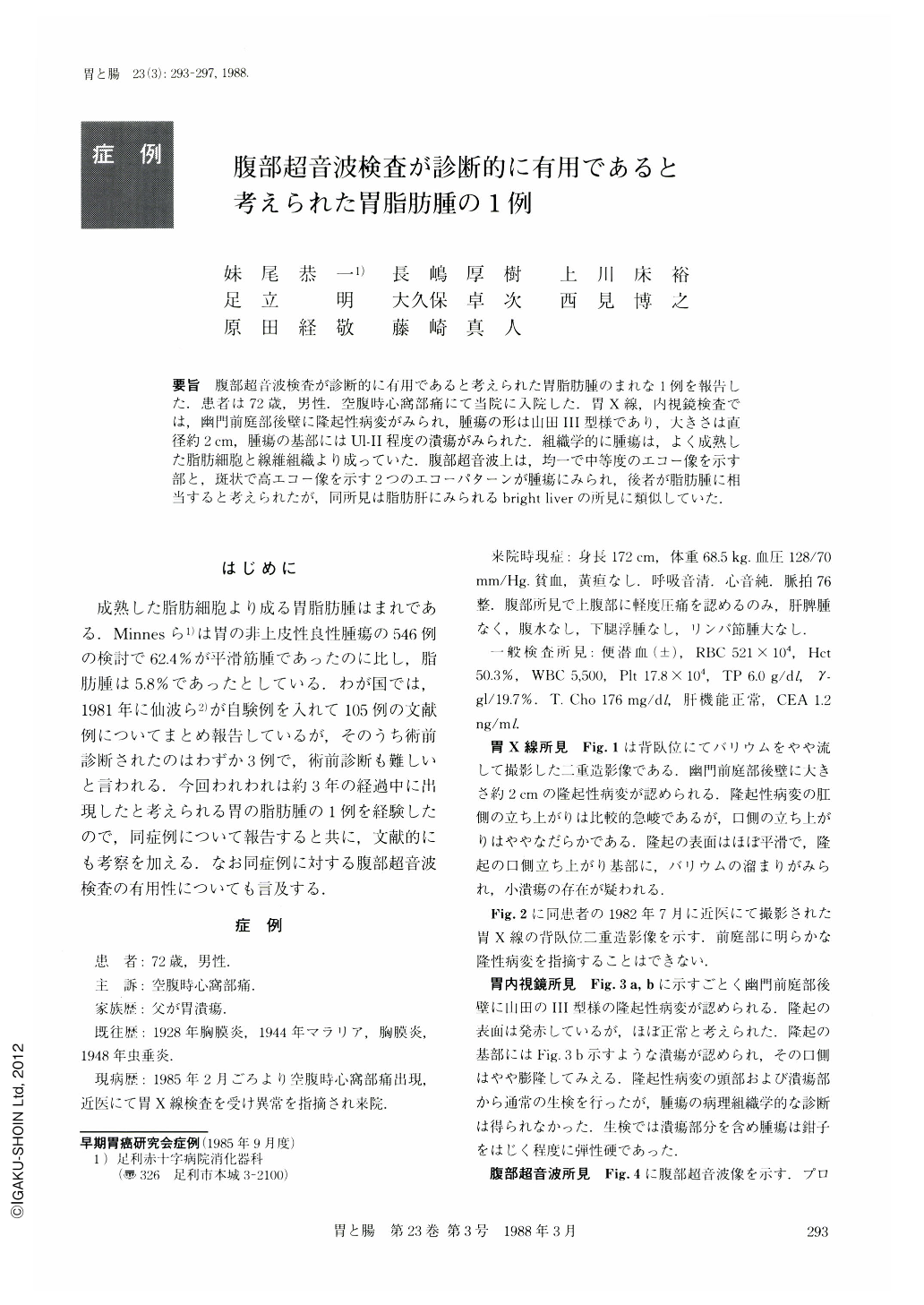
要旨 腹部超音波検査が診断的に有用であると考えられた胃脂肪腫のまれな1例を報告した.患者は72歳,男性.空腹時心窩部痛にて当院に入院した.胃X線,内視鏡検査では,幽門前庭部後壁に隆起性病変がみられ,腫瘍の形は山田Ⅲ型様であり,大きさは直径約2cm,腫瘍の基部にはUl-Ⅱ程度の潰瘍がみられた.組織学的に腫瘍は,よく成熟した脂肪細胞と線維組織より成っていた.腹部超音波上は,均一で中等度のエコー像を示す部と,斑状で高エコー像を示す2つのエコーパターンが腫瘍にみられ,後者が脂肪腫に相当すると考えられたが,同所見は脂肪肝にみられるbright liverの所見に類似していた.
A 72 year-old man was admitted to our hospital with a complaint of hunger-epigastralgia. The roentgenogram and endoscopy showed a protruded lesion on the posterior wall of the antrum. It looked like Yamada type-Ⅲ tumor of about 2 cm in diameter. There was an ulcer (Ul-Ⅱ) at the base of the tumor.
Histologically it was composed of well-matured fat cells and fibrotic tissue. There were two patterns in the abdominal echogram, one being a homogenous and mild echo pattern, and the other a patchy and high echo pattern. The latter, commonly seen in fatty liver, was considered to correspond to the lipoma. Thus, the abdominal echogram may be useful in diagnosing gastric lipoma.
Copyright © 1988, Igaku-Shoin Ltd. All rights reserved.