Japanese
English
- 有料閲覧
- Abstract 文献概要
- 1ページ目 Look Inside
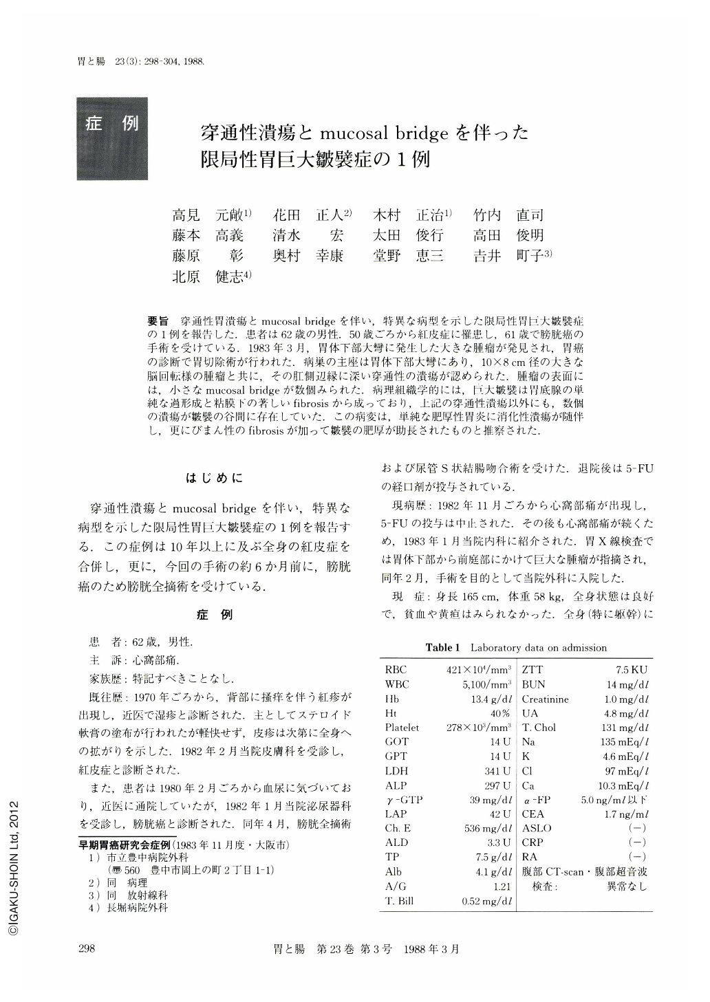
要旨 穿通性胃潰瘍とmucosal bridgeを伴い,特異な病型を示した限局性胃巨大皺襞症の1例を報告した.患者は62歳の男性.50歳ごろから紅皮症に罹患し,61歳で膀胱癌の手術を受けている.1983年3月,胃体下部大彎に発生した大きな腫瘤が発見され,胃癌の診断で胃切除術が行われた.病巣の主座は胃体下部大彎にあり,10×8cm径の大きな脳回転様の腫瘤と共に,その肛側辺縁に深い穿通性の潰瘍が認められた.腫瘤の表面には,小さなmucosal bridgeが数個みられた.病理組織学的には,巨大皺襞は胃底腺の単純な過形成と粘膜下の著しいfibrosisから成っており,上記の穿通性潰瘍以外にも,数個の潰瘍が皺襞の谷間に存在していた.この病変は,単純な肥厚性胃炎に消化性潰瘍が随伴し,更にびまん性のfibrosisが加って皺襞の肥厚が助長されたものと推察された.
A case of localized gastric mucosal hypertrophy simulating neoplasm is reported. A feature unique to this case included an association of radiologically and endoscopically demonstrable gastric ulcer within the lesion.
The patient, a 62 year-old man, was admitted to the hospital in Feb. 1983, complaining of epigastric pain of about three months' duration.
He had been suffering from erythrodermia since ten years prior to the admission and had total cystectomy for an adenocarcinoma of the urinary bladder in April, 1982.
An upper gastrointestinal series revealed a large filling defect compatible with Borrmann type 2 carcinoma. Endoscopy showed localized giant rugal folds on the posterior wall of the lower gastric body associated with a peptic ulcer near the mass of giant rugal folds. Multiple biopsies were negative for malignancy, but the possibility of localized form of scirrous carcinoma or metastatic tumor could not be ruled out.
A Billroth I resection was performed. The resected stomach revealed a large tumor-like mass consisting of giant rugae resembling cerebral convolutions. There was a penetrating peptic ulcer at the distal margin of the mass, which transversed into the giant folds. Peculiar mucosal bridges were noted as well.
Microscopically the giant rugae consisted of simple hyperplasia of fundic mucosa with cystically dilated glands. In addition there was extensive submucosal fibrosis and multiple ulcer scars.
The occurrence of localized hypertrophic gastritis in association with peptic ulcer is rare, but has been reported.
A causal relationship between the two diseases, however, is not clear. In the present case, multiple gastric ulcers were present around and within the region of giant hypertrophic gastritis and were associated with extensive submucosal fibrosis. Therefore, at least, from a morphological standpoint of view, such extensive fibrosis in the submucosal layer may have also contributed in part to the formation of giant rugal folds, a situation similar to that in scirrous carcinoma of the stomach.

Copyright © 1988, Igaku-Shoin Ltd. All rights reserved.


