Japanese
English
- 有料閲覧
PPV購入案内
1,320円 (1,200円+税10%) こちらは、1211ページから1216ページの文献です。購入には医書.jp本会員登録が必要です。
会員登録完了後に改めて上記「購入サイトへ」を選択してください。
または「購入サイトへ」選択後に「購入ログイン」、「新規会員登録」の順に進んでください。
会員登録について詳しくはをご確認ください。
- Abstract 文献概要
- 1ページ目 Look Inside
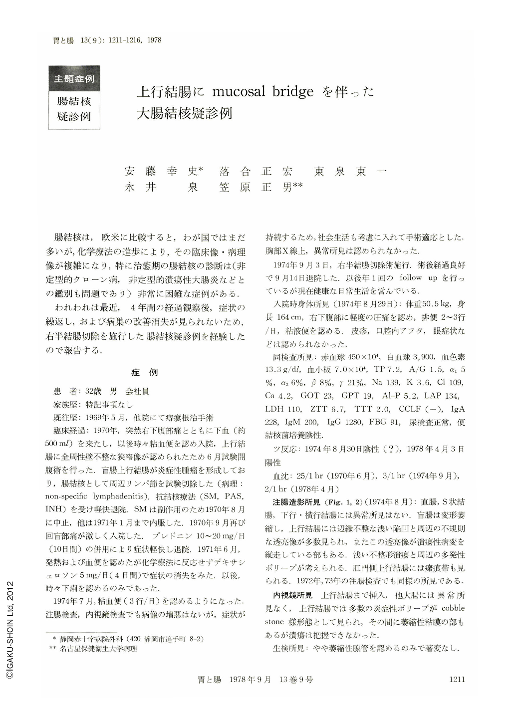
腸結核は,欧米に比較すると,わが国ではまだ多いが,化学療法の進歩により,その臨床像・病理像が複雑になり,特に治癒期の腸結核の診断は(非定型的クローン病,非定型的潰瘍性大腸炎などとの鑑別も問題であり)非常に困難な症例がある.
われわれは最近,4年間の経過観察後,症状の繰返し,および病巣の改善消失が見られないため,右半結腸切除を施行した腸結核疑診例を経験したので報告する.
症 例
患 者:32歳 男 会社員
家族歴:特記事項なし
既往歴:1969年5月,他院にて痔瘻根治手術
A 32-year-old man had a long history of mucoid bloody discharge from the anus and abdominal pain. He had undergone an exploratory laparotomy because of the stenotic lesion of the ascending colon in 1970. Macroscopic diagnosis was tuberculosis of the ascending colon but the regional lymphnodes, were diagnosed pathologically as showing non-specific lymphadenitis. He was treated by antituberculous drugs for six months. During that period he was twice given steroids with good result when the symptoms were aggravated. In August 1974, he had the symptoms of diarrhea with mucoid-bloody discharge and abdominal pain (right lower quadrant).
Barium enema showed deformity of the cecum, ulcer with irregular edge and inflammatory polyps at ascending colon. Barium enema study in 1972 and 1973 had showed similar findings. He was admitted for the resection of the affected region. He had a slight anemia and fecal culture for tubercle bacillus was negative. In September 1974, right hemicolectomy was carried out. Postoperative course was satisfactory and he was discharged on the 11 th day after operation. He has been doing well since discharge.
The operative specimen showed deformity and shortening of the cecum, irregular healed ulcer (5 X 3 cm) and multiple pseudopolyps with mucosal bridge in the ascending colon. Histologically hyperplasia of the lymph follicle and infiltration of lymphocytes were prominent in the mucosal and submucosal layer. Epitheloid cell, giant cell or any specific granuloma was not found. Surface of the polyps consisted of almost normal mucosa but multiple lymphoid tissue existed in the mucosal and submucosal layer. Scar formation of the seromuscular layer was observed at the only one region.
The clinical diagnosis for this case was determined by examining the clinical course, barium enema study, macroscopic appearance and histological study of the resected specimen.
Copyright © 1978, Igaku-Shoin Ltd. All rights reserved.