Japanese
English
- 有料閲覧
- Abstract 文献概要
- 1ページ目 Look Inside
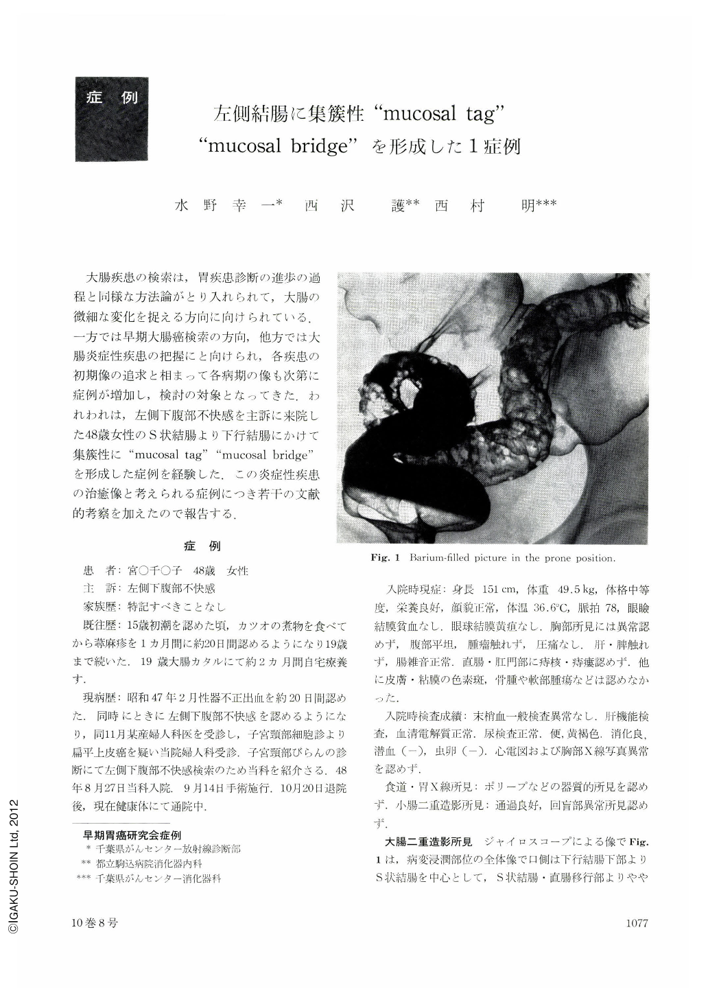
大腸疾患の検索は,胃疾患診断の進歩の過程と同様な方法論がとり入れられて,大腸の微細な変化を捉える方向に向けられている.一方では早期大腸癌検索の方向,他方では大腸炎症性疾患の把握にと向けられ,各疾患の初期像の追求と相まって各病期の像も次第に症例が増加し,検討の対象となってきた.われわれは,左側下腹部不快感を主訴に来院した48歳女性のS状結腸より下行結腸にかけて集簇性に“mucosal tag”“mucosal bridge”を形成した症例を経験した.この炎症性疾患の治癒像と考えられる症例につき若干の文献的考察を加えたので報告する.
This report deals with a case of aggregated “mucosal tag” and “mucosal bridge” presented over a region extending from the upper portion of the rectum to the lower portion of the descending colon. The patient involved was a 48-year-old woman who visited the hospital with pain at the left hypogastric region as chief complaint. In her past history, she suffered from large intestinal catarrh at 19 years of age. She was affected with the symptom mentioned above in February 1972, and visited the out-patient clinic of the hospital in August 1973. Nothing abnormal was revealed by the blood test, biochemical examination, or urinalysis. Occult blood of feces (-). Parasite eggs (-). Contrast medium was injected into the intestine and roentgenological examination performed. Expansion was a little insufficient in a region with the sigmoid colon at its center extending from the lower portion of the descending colon to the junction of the sigmoid colon and the rectum. In this region, the intestinal wall was irregular and some areas of it protruded to form spiculation-like structures. Moreover, spongy “mucosal bridges” irregular in size and various in shape were formed on the mucosal surface. Inflammatory polyps were scattered on the normal mucosa of the descending and transverse colon. Endoscopical examination: Small polyp-like projections were present about 10 cm apart from the annulus haemorrhoidalis. They were denser than those found in the sigmoid colon. They protruded from the mucosal surface to form polyp-like processes round and elliptic in shape and gave rise to hollows with sharp edges. No ulcerative changes were found. Biopsy revealed non-specific inflammatory changes. Nothing abnormal was detected by the X-ray examination of the small intestine. Surgical operation was carried out to excise a region extending from the sigmoid colon to the transverse colon (47 cm long). Pathological changes consisting mostly of “mucosal tag” and “mucosal bridge” were observed in a region extending from the sigmoid colon to the descending colon. No distinct ulcer or erosion was recognized on the mucosal surface. Pathological examination revealed that the tunica propria mucosae was affected with infiltration of a few round cells. The tunica muscularis mucosae was thickened, showing an irregular arrangement of muscle fibers and the deficit of tissue in some areas. The tunica submucosa presented fibrosis accompained by infiltration of a few round cells. The tunica muscularis remained intact. The tunica serosa exhibited inflammatory changes. This case was diagnosed pathologically as that of ulcerative changes extensive and a little shallow which were in the process of healing. Two cases of the same changes have ever been reported in Japan. One of them, as well as the present case, presented these changes in the left colon, and the other in the right colon. All the three cases were free from ulceration.
Copyright © 1975, Igaku-Shoin Ltd. All rights reserved.