Japanese
English
- 有料閲覧
- Abstract 文献概要
- 1ページ目 Look Inside
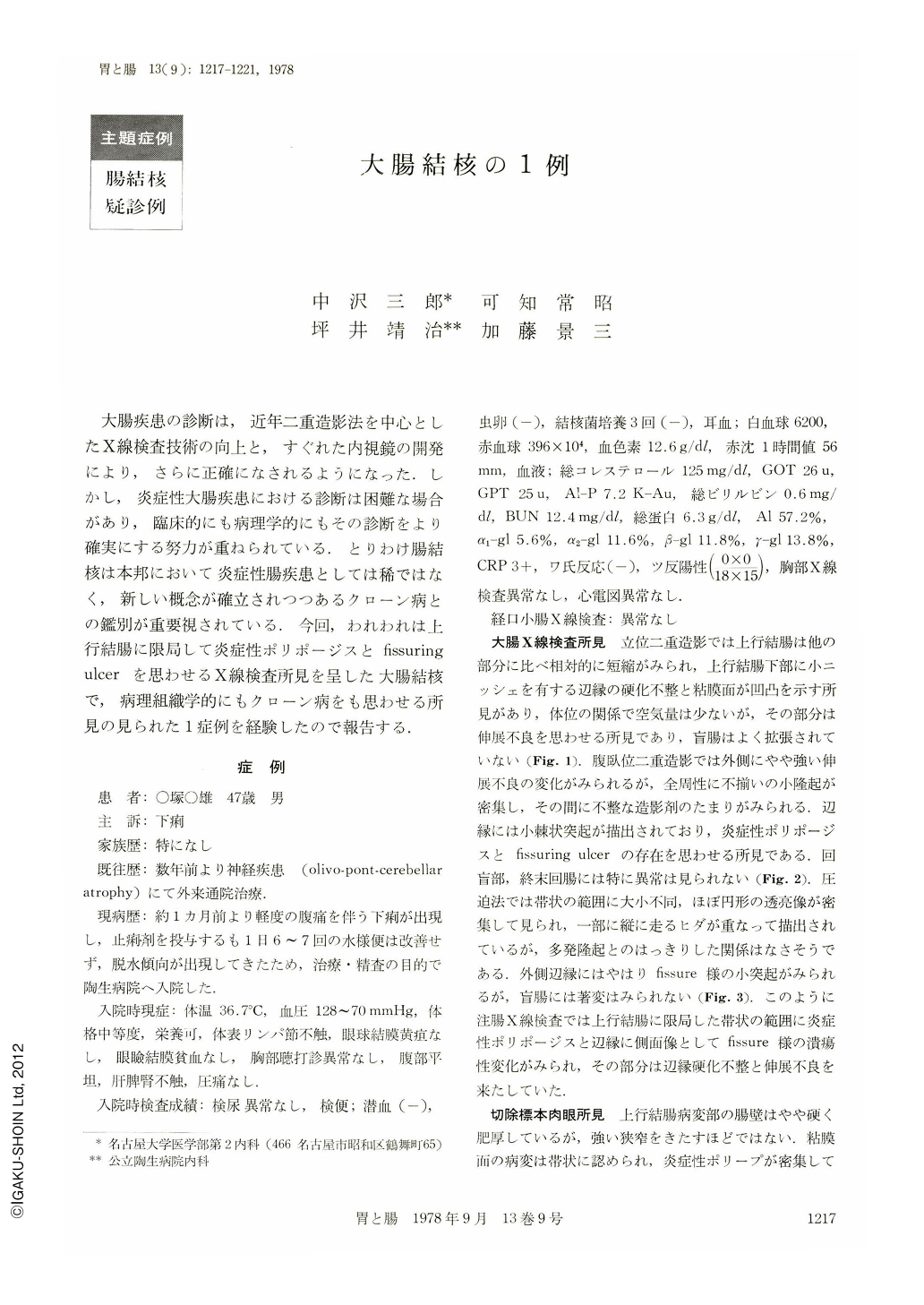
大腸疾患の診断は,近年二重造影法を中心としたX線検査技術の向上と,すぐれた内視鏡の開発により,さらに正確になされるようになった.しかし,炎症性大腸疾患における診断は困難な場合があり,臨床的にも病理学的にもその診断をより確実にする努力が重ねられている.とりわけ腸結核は本邦において炎症性腸疾患としては稀ではなく,新しい概念が確立されつつあるクローン病との鑑別が重要視されている.今回,われわれは上行結腸に限局して炎症性ポリポージスとfissuring ulcerを思わせるX線検査所見を呈した大腸結核で,病理組織学的にもクローン病をも思わせる所見の見られた1症例を経験したので報告する.
症 例
患 者:○塚○雄 47歳 男
主 訴:下痢
家族歴:特になし
既往歴:数年前より神経疾患(olivo-pont-cerebellar atrophy)にて外来通院治療.
現病歴:約1カ月前より軽度の腹痛を伴う下痢が出現し,止痢剤を投与するも1日6~7回の水様便は改善せず,脱水傾向が出現してきたため,治療・精査の目的で陶生病院へ入院した.
A 47 years old man was admitted with complaints of diarrhea and mild abdominal pain. He had a history of olivo-ponto-cerebellar atrophy which he had experienced several years previously.
Roentgenographic examination showed shortening of the ascending colon with inflammatory polyposis and a fissuring ulcer, and slight narrowing.
Resected specimen showed thickening of the bowel wall, inflammatory polyposis and multiple erosions. Microscopy indicated ulcers extending down to the submucosa. Tuberculous granulomas were observed in the submucosal layer, and a few non-caseating granulomas were recognized.
Roentgenographic findings suggested either Crohn's disease or tuberculosis of the ascending colon. Pathologically it was diagnosed as colonic tuberculosis.

Copyright © 1978, Igaku-Shoin Ltd. All rights reserved.


