Japanese
English
- 有料閲覧
PPV購入案内
1,320円 (1,200円+税10%) こちらは、671ページから676ページの文献です。購入には医書.jp本会員登録が必要です。
会員登録完了後に改めて上記「購入サイトへ」を選択してください。
または「購入サイトへ」選択後に「購入ログイン」、「新規会員登録」の順に進んでください。
会員登録について詳しくはをご確認ください。
- Abstract 文献概要
- 1ページ目 Look Inside
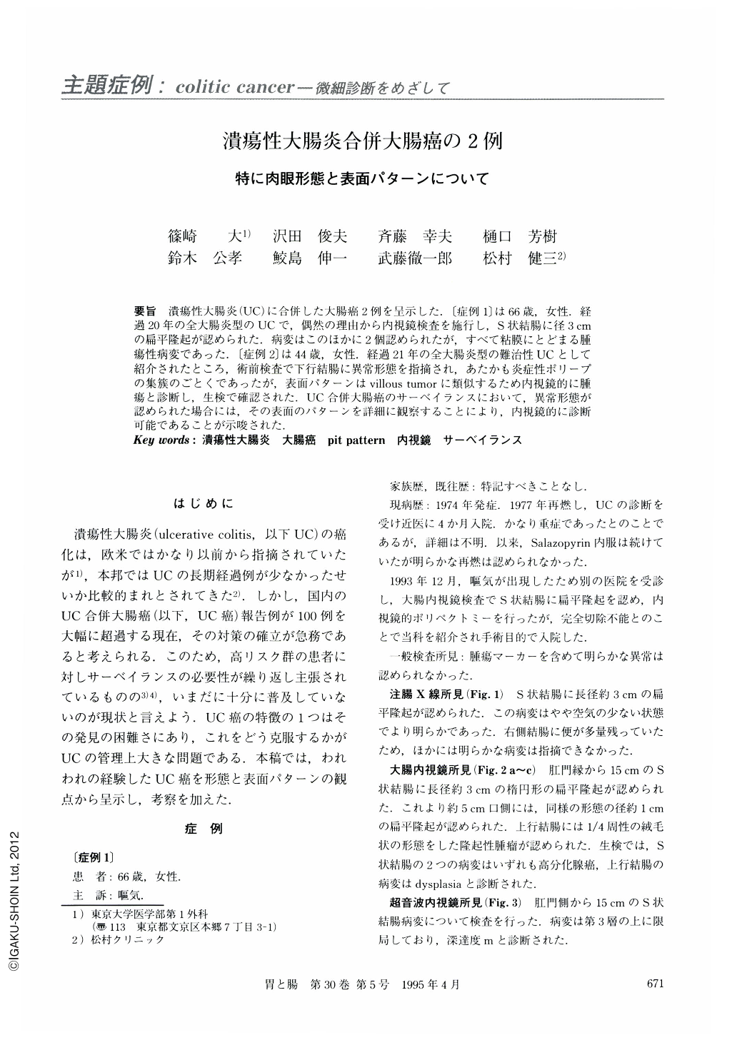
要旨 潰瘍性大腸炎(UC)に合併した大腸癌2例を呈示した.〔症例1〕は66歳,女性.経過20年の全大腸炎型のUCで,偶然の理由から内視鏡検査を施行し,S状結腸に径3cmの扁平隆起が認められた.病変はこのほかに2個認められたが,すべて粘膜にとどまる腫瘍性病変であった.〔症例2〕は44歳,女性.経過21年の全大腸炎型の難治性UCとして紹介されたところ,術前検査で下行結腸に異常形態を指摘され,あたかも炎症性ポリープの集簇のごとくであったが,表面パターンはvillous tumorに類似するため内視鏡的に腫瘍と診断し,生検で確認された.UC合併大腸癌のサーベイランスにおいて,異常形態が認められた場合には,その表面のパターンを詳細に観察することにより,内視鏡的に診断可能であることが示唆された.
Two cases of colon cancer associated with ulcerative colitis were presented. 〔Case 1〕 A 66-year-old female with a 20-year history of ulcerative colitis. Her entire colon was involved. Three lesions were detected in her colon : one was a flat elevation with a larger diameter of 3 cm. All three lesions were neoplastic and localized in the mucosa. Surface pit pattern of the lesions on the resected specimen appeared to be similar to colorectal neoplasms. 〔Case 2〕 A 44-year-old female, with a 21-year history of extensive colitis. On endoscopy, a strange macroscopic configuration like a forest of inflammatory polyps was observed. Its surface pattern was quite similar to that of villous tumor, so we diagnosed this lesion endoscopically as neoplastic, which was confirmed by histology. It is important in colonoscopic surveillance for colitic cancer to observe surface pattern, when visible or detectable lesions are found.
Copyright © 1995, Igaku-Shoin Ltd. All rights reserved.