Japanese
English
- 有料閲覧
- Abstract 文献概要
- 1ページ目 Look Inside
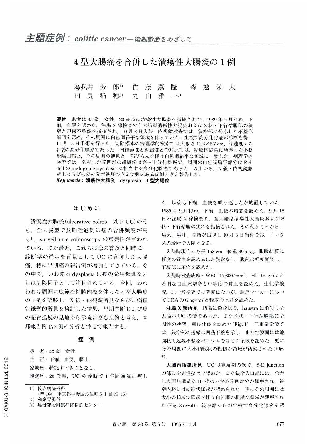
要旨 患者は43歳,女性.20歳時に潰瘍性大腸炎を指摘された.1989年9月初め,下痢,血便を認めた.注腸X線検査で全大腸型潰瘍性大腸炎およびS状・下行結腸部の狭窄と辺縁不整像を指摘され,10月3日入院.内視鏡検査では,狭窄部に発赤した不整形陥凹を認め,その周囲に白色調扁平な領域を伴っていた.生検で高分化腺癌の診断を得,11月15日手術を行った.切除標本の病理学的検索では大きさ11.3×6.7cm,深達度sの4型の高分化腺癌であった.内視鏡像と組織像との対比では,粘膜内癌巣は発赤した不整形陥凹部と,その周囲の褪色と一部びらんを伴う白色調扁平な領域に一致した.病理学的検索では,発赤した陥凹部の組織像は高~中分化腺癌で,周囲の白色調扁平部分はRiddellのhigh-rade dysplasiaに相当する高分化腺癌であった.以上から,X線・内視鏡診断上ならびに癌の発育進展のうえで興味ある症例と考え報告した.
The patient was a 43-year-old woman. She was diagnosed as having ulcerative colitis at the age of 20. Early in September, 1989, she visited a neighborhood clinic because of exacerbating diarrhea and melena and was found, by barium enema x-ray, to have ulcerative colitis of the entire colonic type and stenosis of the descending colon to the sigmoid colon. Because ileus occurred later, she was admitted to our hospital on October 3. A barium enema x-ray study revealed colon wall rigidity and an area of stenosis with an irregular margin. An endoscopic study showed that the site of stenosis had an irregular depression with redness, surface irregularities, and granular elevations. A diagnosis of well differentiated adenocarcinoma was made based on biopsy findings so that sigmoidectomy was performed on November 15, the same year. When the histologic section of resected tissue was examined pathologically, the lesion was found to be a well differentiated type 4 adenocarcinoma 11.3×6.7 cm in size infiltrating to the serosa. When pathologic findings were compared with the endoscopic picture, the intramucosal carcinoma was consistent with the depressed area with redness and with the surrounding whitish flat region with granular elevations of varying size. This case seems to provide very suggestive information which should be helpful for radiologic and endoscopic diagnosis and evaluation of the growth and progression of cancer.

Copyright © 1995, Igaku-Shoin Ltd. All rights reserved.


