Japanese
English
- 有料閲覧
PPV購入案内
1,320円 (1,200円+税10%) こちらは、901ページから909ページの文献です。購入には医書.jp本会員登録が必要です。
会員登録完了後に改めて上記「購入サイトへ」を選択してください。
または「購入サイトへ」選択後に「購入ログイン」、「新規会員登録」の順に進んでください。
会員登録について詳しくはをご確認ください。
- Abstract 文献概要
- 1ページ目 Look Inside
- 参考文献 Reference
- サイト内被引用 Cited by
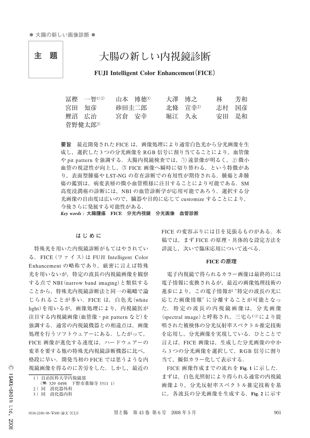
要旨 最近開発されたFICEは,画像処理により通常白色光から分光画像を生成し,選択した3つの分光画像をRGB信号に割り当てることにより,血管像やpit patternを強調する.大腸内視鏡検査では,①遠景像が明るく,②微小血管の視認性が向上し,③FICE画像へ瞬時に切り替わる,という特徴があり,表面型腫瘍やLST-NGの存在診断での有用性が期待される.腫瘍と非腫瘍の鑑別は,病変表層の微小血管模様に注目することにより可能である.SM高度浸潤癌の診断には,NBIの血管診断学が応用可能であろう.選択する分光画像の自由度は広いので,臓器や目的に応じてcustomizeすることにより,今後さらに発展する可能性がある.
Fuji intelligent color enhancement (FICE) is an image-processing apparatus for video endoscopy where 3 spectral images for each wavelength are selected from ordinary endoscopic images and then allocated an RGB signal to visualize the capillary network and pit pattern on the mucosal surface. The FICE image has interesting features, i. e., clear distant view, improved visibility of capillary network and instant access to FICE image. These features imply better detectability of flat adenomas and LST-NG than the conventional plain procedure. It is possible to differentiate neoplastic from non-neoplastic polyps based on the capillary pattern of the polyp surface. Polyp cancers with substantial invasion can be identified from the irregularity of the capillary network. Millions of FICE images can be processed from one ordinary endoscopic image. To obtain a much clearer colonoscopic image, we have to fully customize FICE.
Copyright © 2008, Igaku-Shoin Ltd. All rights reserved.