Japanese
English
- 有料閲覧
PPV購入案内
1,320円 (1,200円+税10%) こちらは、1397ページから1405ページの文献です。購入には医書.jp本会員登録が必要です。
会員登録完了後に改めて上記「購入サイトへ」を選択してください。
または「購入サイトへ」選択後に「購入ログイン」、「新規会員登録」の順に進んでください。
会員登録について詳しくはをご確認ください。
- Abstract 文献概要
- 1ページ目 Look Inside
- 参考文献 Reference
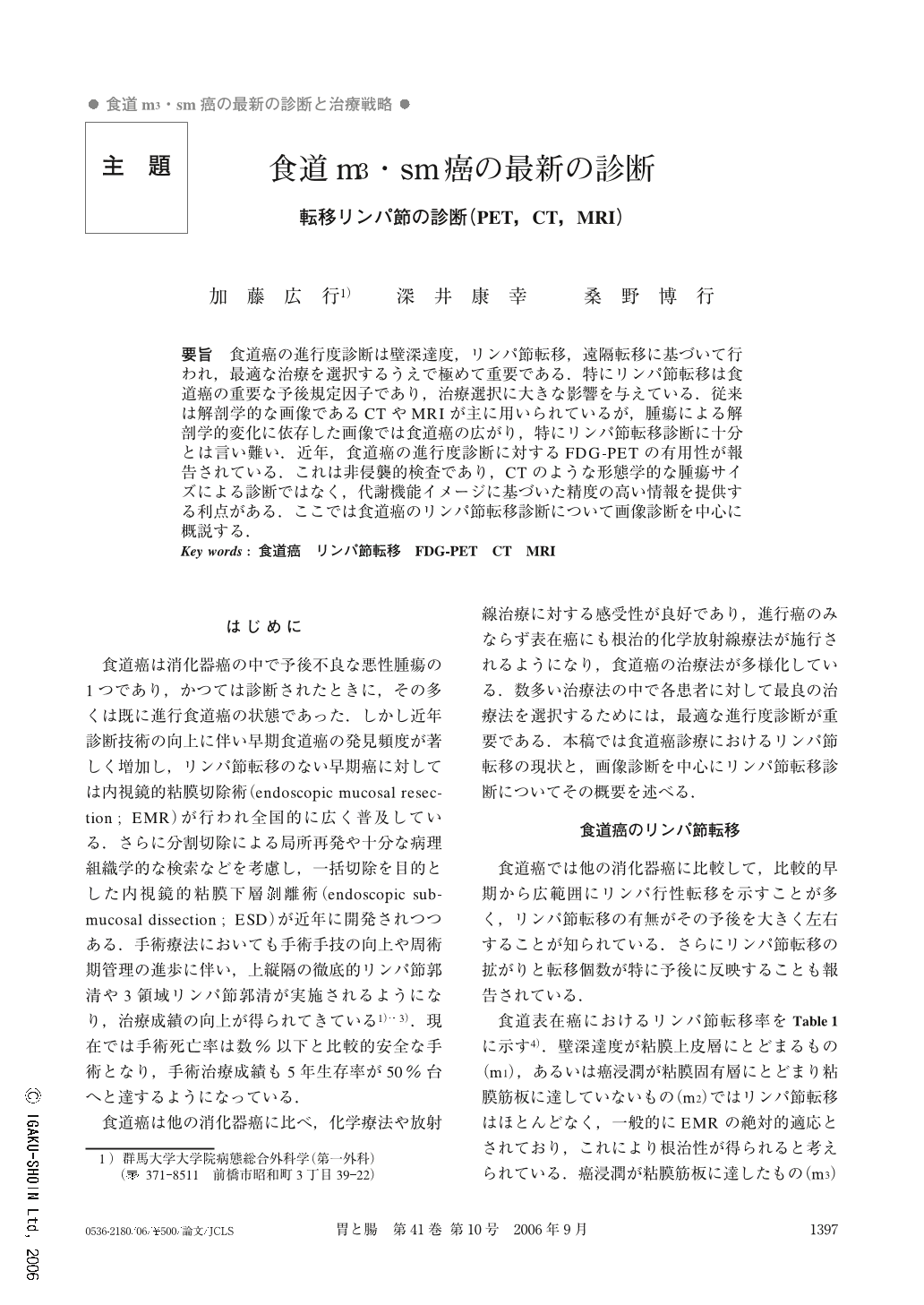
要旨 食道癌の進行度診断は壁深達度,リンパ節転移,遠隔転移に基づいて行われ,最適な治療を選択するうえで極めて重要である.特にリンパ節転移は食道癌の重要な予後規定因子であり,治療選択に大きな影響を与えている.従来は解剖学的な画像であるCTやMRIが主に用いられているが,腫瘍による解剖学的変化に依存した画像では食道癌の広がり,特にリンパ節転移診断に十分とは言い難い.近年,食道癌の進行度診断に対するFDG-PETの有用性が報告されている.これは非侵襲的検査であり,CTのような形態学的な腫瘍サイズによる診断ではなく,代謝機能イメージに基づいた精度の高い情報を提供する利点がある.ここでは食道癌のリンパ節転移診断について画像診断を中心に概説する.
Accurate pre-treatment staging, particularly with regard to the depth of tumor invasion, lymph node involvement, and distant metastases, is vital in determining the most appropriate procedures for the management of esophageal carcinomas. In particular, lymph node staging is an important independent prognostic indicator in esophageal cancer and has a major impact on treatment selection. Conventional anatomical imaging procedures, including computed tomography (CT) and magnetic resonance imaging (MRI), have been available for the pre-treatment staging of esophageal cancer. The imaging methods that rely on detection of the structural changes caused by tumors are inaccurate in determining the extent of esophageal cancer, especially lymph node metastasis. Recent studies have reported on the efficacy of 18F-fluorodeoxyglucose positron emission tomography (FDG-PET) for staging of esophageal carcinoma. FDG-PET imaging is noninvasive and provides qualitatively different information compared with CT imaging, due to its reliance on the metabolic function of tumors rather than tumor size alone. In the current article, we review the performance of main imaging methods in the assessment of lymph node staging of esophageal carcinoma.
Copyright © 2006, Igaku-Shoin Ltd. All rights reserved.