Japanese
English
- 有料閲覧
PPV購入案内
1,320円 (1,200円+税10%) こちらは、1675ページから1678ページの文献です。購入には医書.jp本会員登録が必要です。
会員登録完了後に改めて上記「購入サイトへ」を選択してください。
または「購入サイトへ」選択後に「購入ログイン」、「新規会員登録」の順に進んでください。
会員登録について詳しくはをご確認ください。
- Abstract 文献概要
- 1ページ目 Look Inside
- 参考文献 Reference
- サイト内被引用 Cited by
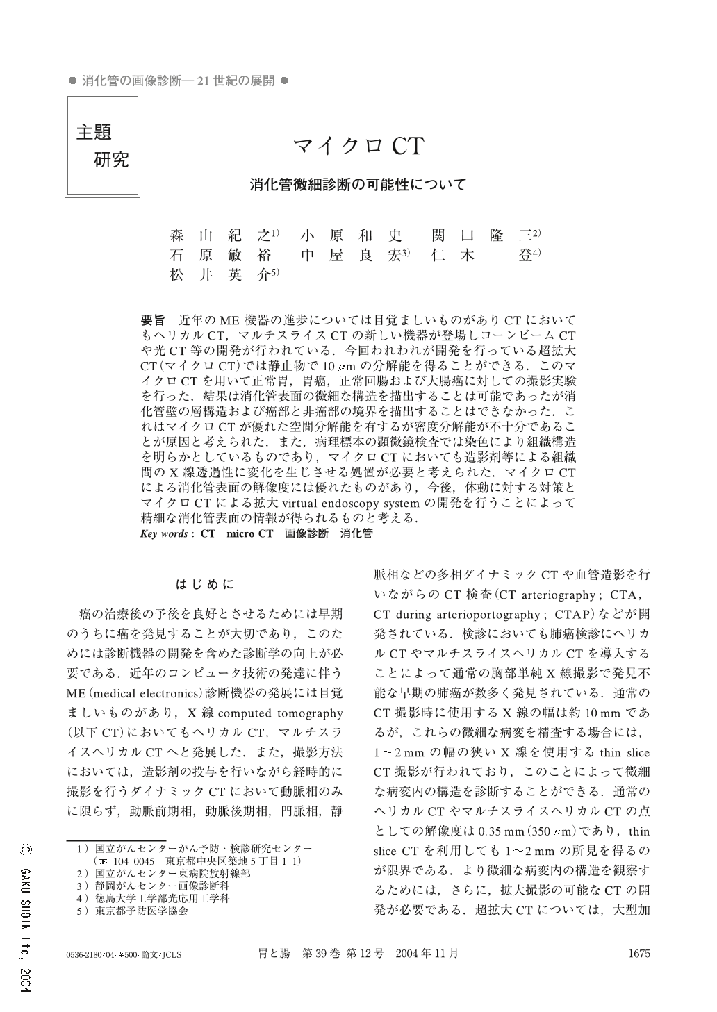
要旨 近年のME機器の進歩については目覚ましいものがありCTにおいてもヘリカルCT,マルチスライスCTの新しい機器が登場しコーンビームCTや光CT等の開発が行われている.今回われわれが開発を行っている超拡大CT(マイクロCT)では静止物で10μmの分解能を得ることができる.このマイクロCTを用いて正常胃,胃癌,正常回腸および大腸癌に対しての撮影実験を行った.結果は消化管表面の微細な構造を描出することは可能であったが消化管壁の層構造および癌部と非癌部の境界を描出することはできなかった.これはマイクロCTが優れた空間分解能を有するが密度分解能が不十分であることが原因と考えられた.また,病理標本の顕微鏡検査では染色により組織構造を明らかとしているものであり,マイクロCTにおいても造影剤等による組織間のX線透過性に変化を生じさせる処置が必要と考えられた.マイクロCTによる消化管表面の解像度には優れたものがあり,今後,体動に対する対策とマイクロCTによる拡大virtual endoscopy systemの開発を行うことによって精細な消化管表面の情報が得られるものと考える.
We are developing a microscopic X-ray computed tomography (microscopic CT) System. The basic components of the microscopic CT system consist of a microfocus X-ray source, a specimen manipulator, and an image intensifier detector coupled to a charge-coupled device (CCD) camera. A standard fan-beam convolution and backprojection algorithm was used to reconstruct the center plane intersecting the X-ray source. The main advance of the system is to obtain high resolution which ranges to 10μm. In this paper we report our preliminary studies carried out with the microscopic CT for imaging surgically resected, inflated and fixed lung specimens of two peripheral pulmonary adenocarcinoma cases (one or them combined with pulmonary emphysema), stomach specimens of 2 cases (normal stomach and type 4 gastric cancer) and normal ileum. Experimental results reveal the micro structure of lung tissues, such as alveolar walls, interlobular septa, bronchioles, and intralesional architectures. However microscopic CT dose not reveal the structure of the gastric wall. Microscopic CT has good space resolution, but density resolution is poor due to the insufficiency of X-ray dose. Judging from the results so far, the micro CT system is expected to have interesting potentials for experimental and clinical investigation. There are many hurdles to be cleared before we can use this microscopic CT clinically.
1) Research Center for Cancer Prevention and Screening, National Cancer Center Hospital, Tokyo
2) Department of Radiology, National Cancer Center East, Kashiwa, Japan
3) Department of Radiology, Shizuoka Cancer Center,Sizuoka, Japan
Copyright © 2004, Igaku-Shoin Ltd. All rights reserved.