Japanese
English
- 有料閲覧
- Abstract 文献概要
- 1ページ目 Look Inside
- 参考文献 Reference
要旨
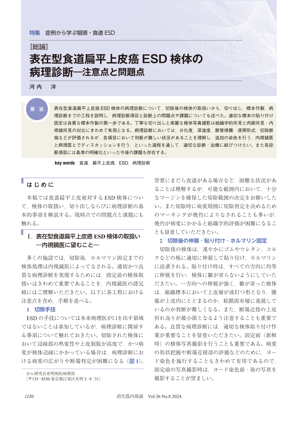
表在型食道扁平上皮癌ESD検体の病理診断について,切除後の検体の取扱いから,切り出し,標本作製,病理診断までの工程を説明し,病理診断項目と診断上の問題点や課題についても述べた。適切な標本の貼り付け固定は良質な標本作製の第一歩である。丁寧な切り出しと美麗な検体写真撮影は組織学的所見と肉眼所見・内視鏡所見の対比にきわめて有用となる。病理診断においては,分化度,深達度,脈管侵襲,浸潤形式,切除断端などが評価されるが,各項目において判断が難しい状況があることを理解し,追加の染色を行う,内視鏡医と病理医とでディスカッションを行う,といった過程を通して,適切な診断・治療に結びつけたい。また各診断項目には基準の明確化といった今後の課題も存在する。
This study outlines the pathological diagnosis of superficial esophageal squamous cell carcinoma in endoscopic submucosal dissection specimens, detailing the processes from post-resection specimen handling, sectioning, and slide preparation to pathological diagnosis. It also discusses the diagnostic criteria and the challenges and issues encountered in diagnosis. Adequate stretching and fixation of specimens by endoscopists are the first steps toward high-quality slide preparation. Careful sectioning and taking clear photographs of the specimens are extremely useful for correlating histological findings with macroscopic findings and endoscopic observations. In pathological diagnosis, factors such as tumor differentiation, depth of invasion, lymphovascular invasion, pattern of infiltration, and resection margins are evaluated. However, it is important to recognize that these factors may present difficult situations requiring additional staining and discussions between endoscopists and pathologists to achieve adequate diagnosis and treatment. There are future challenges, such as the need to clarify the standards for each diagnostic criterion.

© tokyo-igakusha.co.jp. All right reserved.


