Japanese
English
- 有料閲覧
- Abstract 文献概要
- 1ページ目 Look Inside
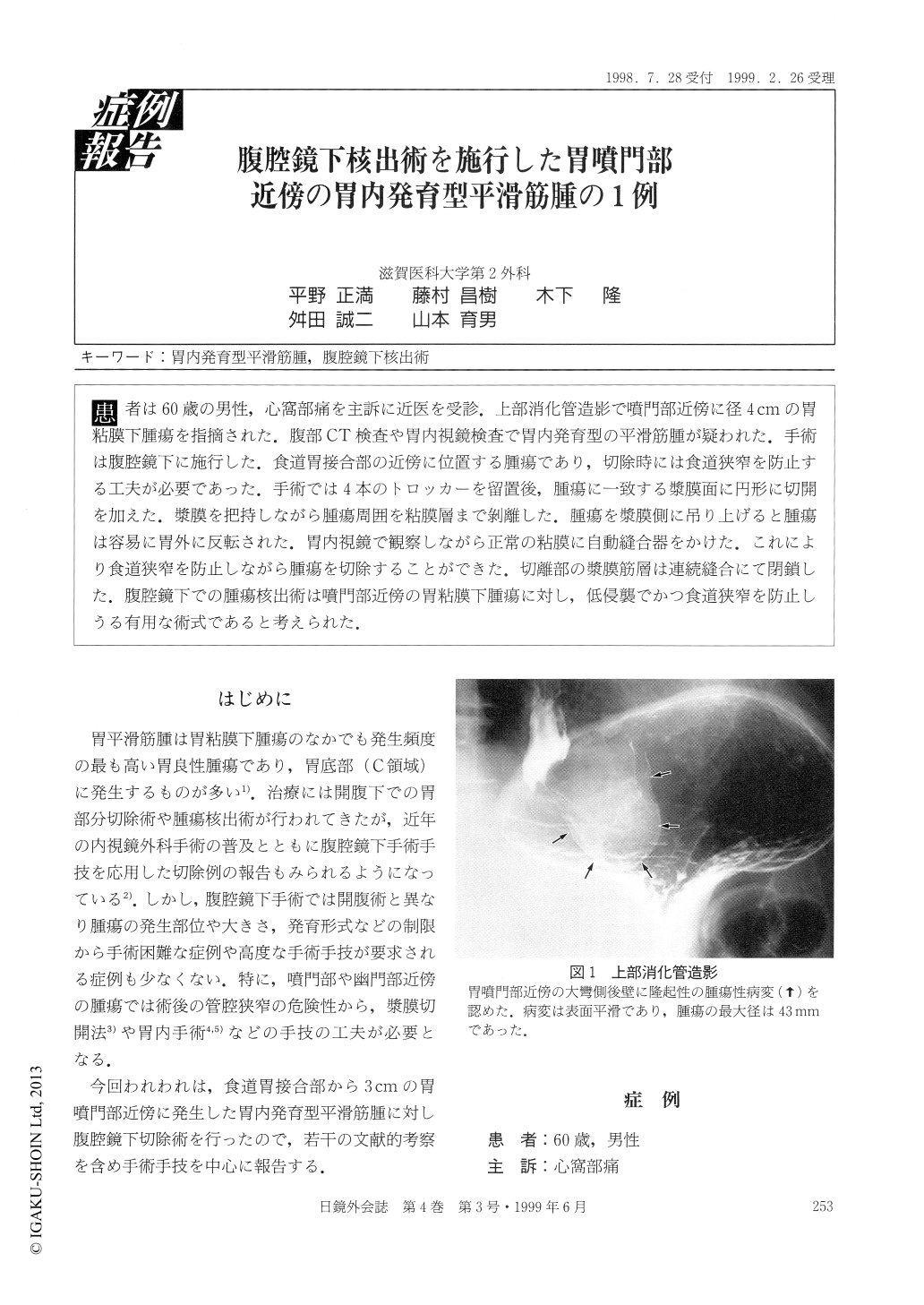
患者は60歳の男性,心窩部痛を主訴に近医を受診.上部消化管造影で噴門部近傍に径4cmの胃粘膜下腫瘍を指摘された.腹部CT検査や胃内視鏡検査で胃内発育型の平滑筋腫が疑われた.手術は腹腔鏡下に施行した.食道胃接合部の近傍に位置する腫瘍であり,切除時には食道狭窄を防止する工夫が必要であった.手術では4本のトロッカーを留置後,腫瘍に一致する漿膜面に円形に切開を加えた.漿膜を把持しながら腫瘍周囲を粘膜層まで剥離した.腫瘍を漿膜側に吊り上げると腫瘍は容易に胃外に反転された.胃内視鏡で観察しながら正常の粘膜に自動縫合器をかけた.これにより食道狭窄を防止しながら腫瘍を切除することができた.切離部の漿膜筋層は連続縫合にて閉鎖した.腹腔鏡下での腫瘍核出術は噴門部近傍の胃粘膜下腫瘍に対し,低侵襲でかつ食道狭窄を防止しうる有用な術式であると考えられた.
A 60-year-old male visited the clinic with a symptom of epigastralgia. A barium upper gastrointestinal series showed a submucosal tumor 4 cm in diameter, on the greater curvature near the cardia. Abdominal CT scan and gastrofiberscopic examination suggested a leiomyoma of the stomach. The patient was admitted to our hospital and a resection of the tumor was performed laparoscopically. Because the tumor was located close to the esophagogastric junction, prevention of stenosis of the esophageal lumen was required.

Copyright © 1999, JAPAN SOCIETY FOR ENDOSCOPIC SURGERY All rights reserved.


