Japanese
English
- 有料閲覧
- Abstract 文献概要
- 1ページ目 Look Inside
I.はじめに
口蓋扁桃にポリープ形成をきたす疾患はそれ程多くはない。代表的疾患は振子様扁桃とよばれるもので,扁桃組織からなる腫瘤が有茎性,ポリープ様に突出する場合である。
今回筆者らは臨床的に外観から振子様扁桃と診断し,切除術を施行し,病理組織学的に検討したところ,極めて稀なlymphangiectatic fibrouspolypと診断された3症例を経験したので,文献的考察を加えて報告する。
Three cases of lymphangiectatic fibrous polyp in the palatine tonsil are reported.
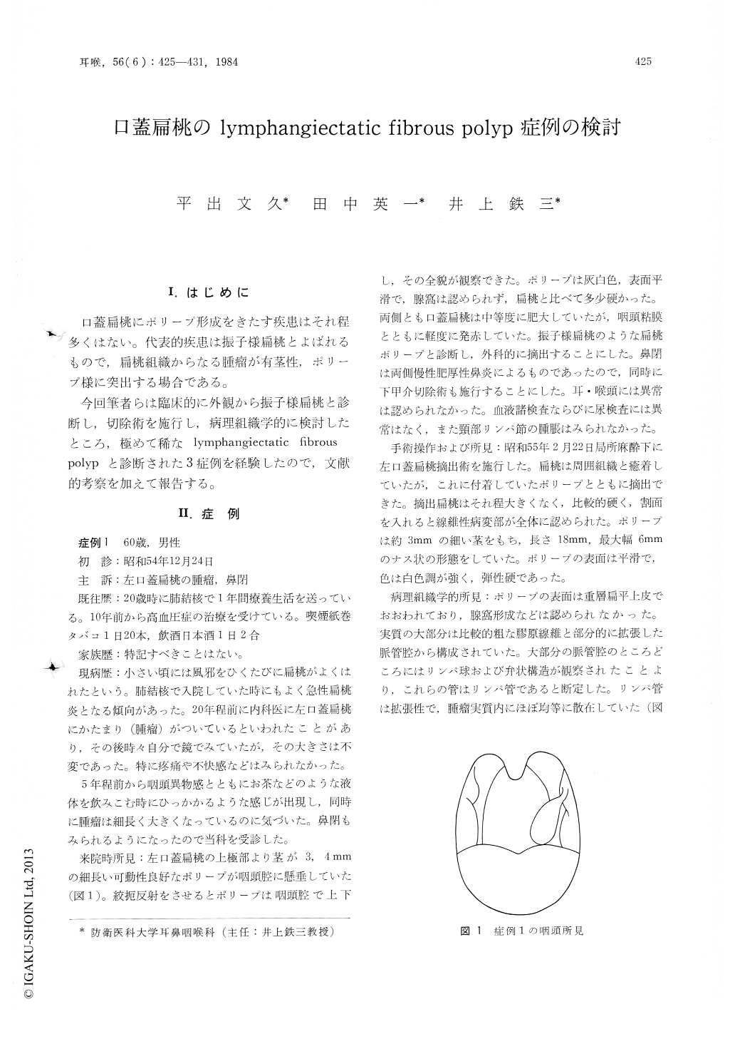
Case 1, a man aged 60, presented in the clinic with complaints of presence of the tonsillar mass and foreign body sensation in the throat, which had been noticed for approximately 20 years. Visual examination revealed that an elongated oval-shaped polyp was hanging from the upper pole of the left palatine tonsil down to the pharynx. The polyp was pinkish-white. Its surface was entirely smooth and not cryptic. It was provisionally diagnosed as a pendulous tonsil. The polyp was removed along with the tonsil under local anesthesia. It was histopathologically diagnosed as lymphangiectatic fibrous polyp of the tonsil.
Case 2, a man aged 44, was referred to the clinic by an otolaryngologist because of presence of a tumorous mass in the palatine tonsil. On examination, there was a large rather pedunculated growth arising from the middle to lower part of the left palatine tonsil and extending beyond the midline of the pharynx. Its surface was almost smooth and not cryptic. A clinical diagnosis of a pendulous tonsil was made. The excised polyp was histopathologically diagnosed of lymphangiectatic fibrous polyp of the tonsil.
Case 3, a girl aged 13, had noted a mass on her right palatine tonsil approximately 10 years before she came to the clinic. In recent years the mass gradually increased in size, but produced no symptoms, except for that she inadvertently partially swallow it. Visual examination revealed a large whitish pedunculated mass growing from the crypt of the upper part of the tonsil. On palpation, the polypous mass was smooth and soft. A clinical diagnosis was made of a pendulous tonsil. The polyp was removed by sectioning its pedicle. Its histological section showed a typical lymphangiectatic fibrous polyp originating from the palatine tonsil.
Copyright © 1984, Igaku-Shoin Ltd. All rights reserved.