Japanese
English
- 有料閲覧
- Abstract 文献概要
- 1ページ目 Look Inside
Ⅰ.はじめに
一般に疾病罹患部位の写真撮影は,最も客観的な所見を記録し得る手段であり,診断の結果を他に伝達し,或は発表する目的に古来各科に於て用いられてきた。しかもそれが我が科領域の如く,罹患部位が患者自身には見えず,我々医師にのみ見えるのであるから,写真は患者に対しても大きな説得力を持つていることは明らかである。
しかし,元来耳鼻咽喉科領域に於ける写真撮影は,その対象となる部位が多岐にわたり,しかも内腔の形態条件が各々異るという悪条件により,今日尚普及する段階に到つてはいない。
Photography against each cavity in our fi-eld is not so popular way, because customar-illy special camera are so expensive and so complicated when clinician want to use them.
Further, endoscopic optical system of their camera is not ideal one for anastigmatic ph-otograph, because the lens is so small.
Retinal camera is made for photography through the pupil.
This condition can apply to another small cavity, but traditional retinal cameras were made for use on the table top.
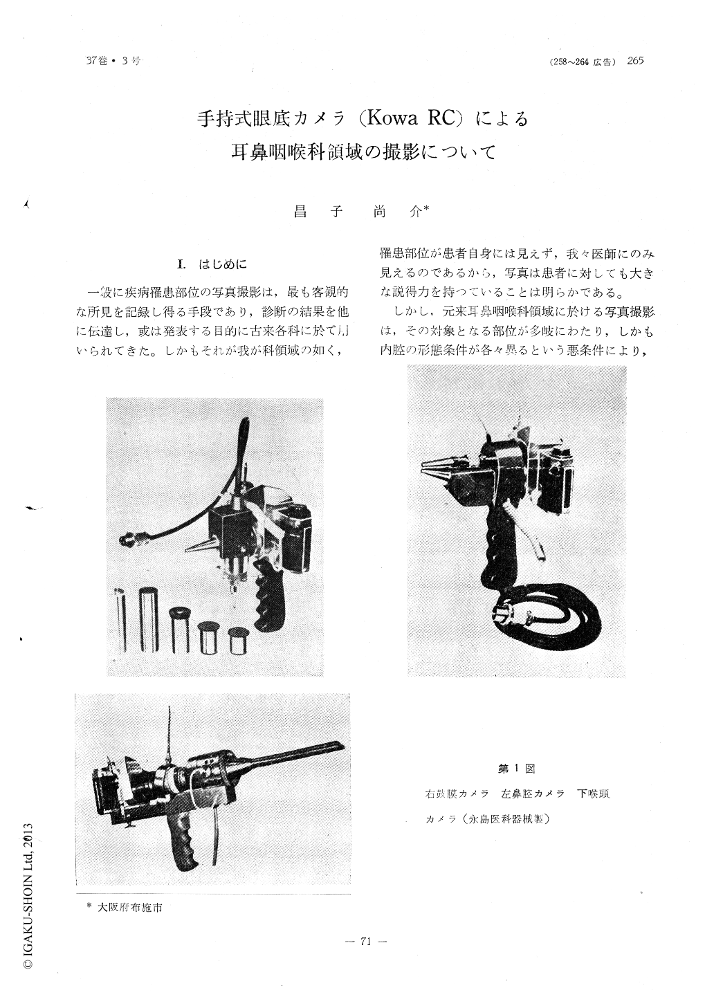
The another took pictures of tympanic-me-mbranes, nasal-, pharyngeal cavities, vocal cords and operations fields with recently pro-duced hand-hold retinal camera.
These photography is very simple and very speedy.
In the use of this, usefulness and chance of our photography can result wide and fre-quent.

Copyright © 1965, Igaku-Shoin Ltd. All rights reserved.


