Japanese
English
- 有料閲覧
- Abstract 文献概要
- 1ページ目 Look Inside
I.はじめに
海綿状血管腫は体のあらゆる部位に生じる血管奇形の一種であり,脳実質内には人口の約O.4〜0.5%に生じると報告されている2,25).近年,MRIの普及と撮像法の発達に伴い,家族性に発症する報告が散見される.われわれは父子に多発性の脳内海綿状血管腫を認めた症例を経験したので,日本人での報告を中心に文献的考察を加え報告する.また,家族性の海綿状血管腫に対しては放射線治療が重要な治療法と考えられ,併せて考察を加える.
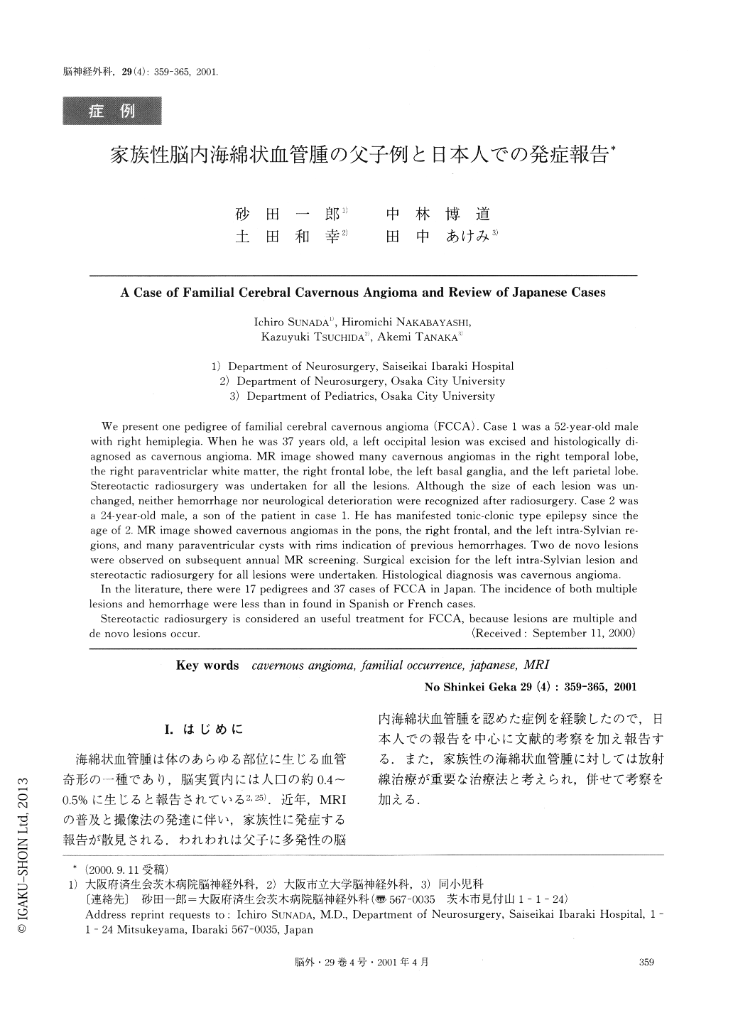
We present one pedigree of familial cerebral cavernous angioma (FCCA). Case 1 was a 52-year-old male with rlght hemiplegia. When he was 37 years old, a left occipital lesion was excised and histologlcally di-agnosed as cavernous angioma. MR image showed many cavernous angiomas in the right temporal lobe, the right paraventriclar white matter, the right frontal lobe, the left basal ganglia, and the left parietal lobe. Stereotactic radiosurgery was undertaken for all the lesions. Although the size of each lesion was un-changed, neither hemorrhage nor neurological deterioration were recognized after radiosurgery.

Copyright © 2001, Igaku-Shoin Ltd. All rights reserved.


