Japanese
English
- 有料閲覧
PPV購入案内
1,320円 (1,200円+税10%) こちらは、53ページから58ページの文献です。購入には医書.jp本会員登録が必要です。
会員登録完了後に改めて上記「購入サイトへ」を選択してください。
または「購入サイトへ」選択後に「購入ログイン」、「新規会員登録」の順に進んでください。
会員登録について詳しくはをご確認ください。
- Abstract 文献概要
- 1ページ目 Look Inside
- 参考文献 Reference
Ⅰ.はじめに
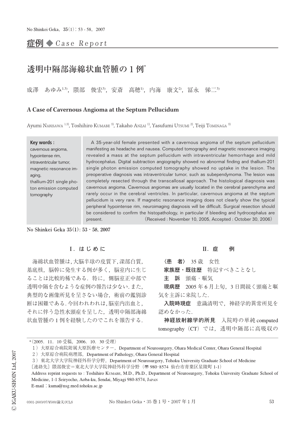
海綿状血管腫は,大脳半球の皮質下,深部白質,基底核,脳幹に発生する例が多く,脳室内に生じることは比較的稀である.特に,側脳室正中部で透明中隔を含むような症例の報告は少ない.また,典型的な画像所見を呈さない場合,術前の鑑別診断は困難である.今回われわれは,脳室内出血と,それに伴う急性水頭症を呈した,透明中隔部海綿状血管腫の1例を経験したのでこれを報告する.
A 35-year-old female presented with a cavernous angioma of the septum pellucidum manifesting as headache and nausea. Computed tomography and magnetic resonance imaging revealed a mass at the septum pellucidum with intraventricular hemorrhage and mild hydrocephalus. Digital subtraction angiography showed no abnormal finding and thallium-201 single photon emission computed tomography showed no uptake in the lesion. The preoperative diagnosis was intraventricular tumor, such as subependymoma. The lesion was completely resected through the transcallosal approach. The histological diagnosis was cavernous angioma. Cavernous angiomas are usually located in the cerebral parenchyma and rarely occur in the cerebral ventricles. In particular, cavernous angioma at the septum pellucidum is very rare. If magnetic resonance imaging does not clearly show the typical peripheral hypointense rim, neuroimaging diagnosis will be difficult. Surgical resection should be considered to confirm the histopathology, in particular if bleeding and hydrocephalus are present.
Copyright © 2007, Igaku-Shoin Ltd. All rights reserved.