Japanese
English
- 有料閲覧
PPV購入案内
1,320円 (1,200円+税10%) こちらは、429ページから434ページの文献です。購入には医書.jp本会員登録が必要です。
会員登録完了後に改めて上記「購入サイトへ」を選択してください。
または「購入サイトへ」選択後に「購入ログイン」、「新規会員登録」の順に進んでください。
会員登録について詳しくはをご確認ください。
- Abstract 文献概要
- 1ページ目 Look Inside
- 参考文献 Reference
Ⅰ.はじめに
Okadaら11)により初めて報告され,Perryら12)により命名された硬膜海綿状血管腫は,頭頂円蓋部硬膜下腔を好発部位とする髄外血管腫のことである.これまでに10例の報告があり,病理学的には髄内血管腫と同等だが,髄膜腫との鑑別がしばしば困難であるなど臨床的には異なる疾患であると考えられている5,7,11,12,15-17,19,20).
われわれの経験した硬膜海綿状血管腫の1例を,文献的考察を加え報告する.
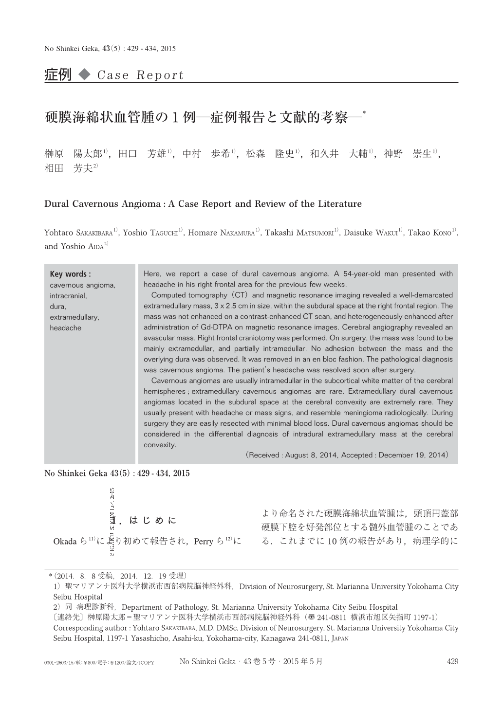
Here, we report a case of dural cavernous angioma. A 54-year-old man presented with headache in his right frontal area for the previous few weeks.
Computed tomography(CT)and magnetic resonance imaging revealed a well-demarcated extramedullary mass, 3 x 2.5cm in size, within the subdural space at the right frontal region. The mass was not enhanced on a contrast-enhanced CT scan, and heterogeneously enhanced after administration of Gd-DTPA on magnetic resonance images. Cerebral angiography revealed an avascular mass. Right frontal craniotomy was performed. On surgery, the mass was found to be mainly extramedullar, and partially intramedullar. No adhesion between the mass and the overlying dura was observed. It was removed in an en bloc fashion. The pathological diagnosis was cavernous angioma. The patient's headache was resolved soon after surgery.
Cavernous angiomas are usually intramedullar in the subcortical white matter of the cerebral hemispheres;extramedullary cavernous angiomas are rare. Extramedullary dural cavernous angiomas located in the subdural space at the cerebral convexity are extremely rare. They usually present with headache or mass signs, and resemble meningioma radiologically. During surgery they are easily resected with minimal blood loss. Dural cavernous angiomas should be considered in the differential diagnosis of intradural extramedullary mass at the cerebral convexity.
Copyright © 2015, Igaku-Shoin Ltd. All rights reserved.