Japanese
English
- 有料閲覧
- Abstract 文献概要
- 1ページ目 Look Inside
I.はじめに
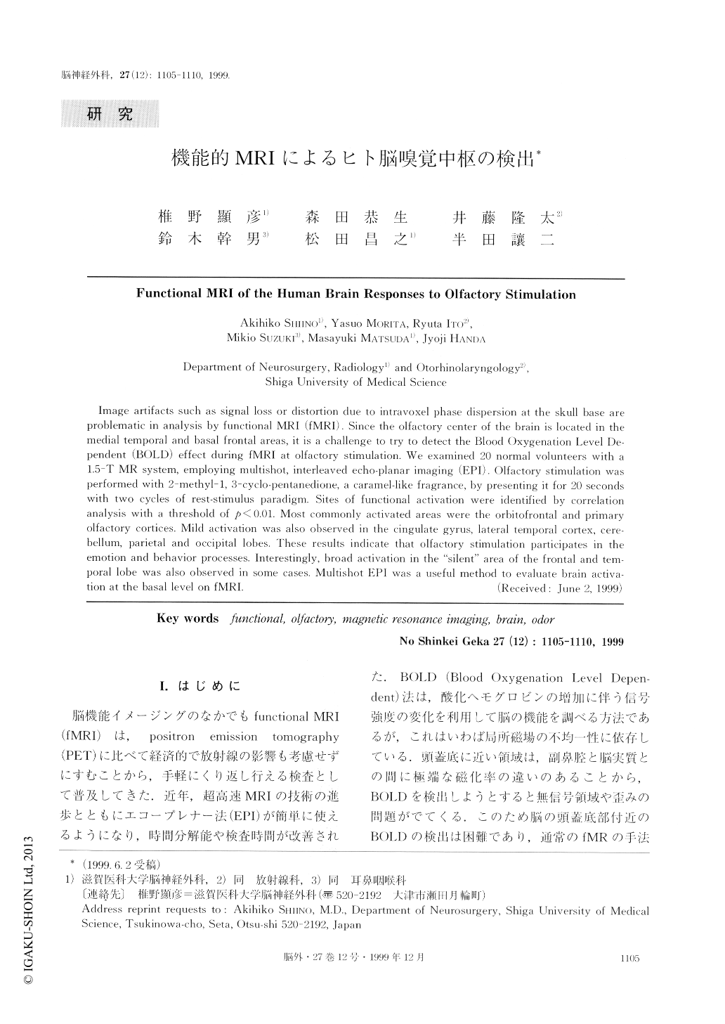
脳機能イメージングのなかでもfunctiomal MRI(fMRI)は,positron emission tomography(PET)に比べて経済的で放射線の影響も考慮せずにすむことから,手軽にくり返し行える検査として普及してきた.近年,超高速MRIの技術の進歩とともにエコープレナー法(EPI)が簡単に使えるようになり,時間分解能や検査時間が改善された.BOLD(Blood Oxygenation Level Depen-dent)法は,酸化ヘモグロビンの増加に伴う信号強度の変化を利用して脳の機能を調べる方法であるが,これはいわば局所磁場の不均一性に依存している.頭蓋底に近い領域は,副鼻腔と脳実質との間に極端な磁化率の違いのあることから,BOLDを検出しようとすると無信号領域や歪みの問題がでてくる.このため脳の頭蓋底部付近のBOLDの検出は困難であり,通常のfMRの手法では賦活領域を描出できない.今回われわれは,このsusceptibilityの問題を克服すべく嗅覚刺激に対する中枢の反応をfMRを用いて検討したので報告する.
Image artifacts such as signal loss or distortion due to intravoxel phase dispersion at the skull base areproblematic in analysis by functional MRI (fMRI). Since the olfactory center of the brain is located in themedial temporal and basal frontal areas, it is a challenge to try to detect the Blood Oxygenation Level De-pendent (BOLD) effect during fMRI at olfactory stimulation. We examined 20 normal volunteers with a1.5-T MR system, employing multishot, interleaved echo-planar imaging (EPI). Olfactory stimulation wasperformed with 2-methyl-1, 3-cyclo-pentanedione, a caramel-like fragrance, by presenting it for 20 secondswith two cycles of rest-stimulus paradigm. Sites of functional activation were identified by correlationanalysis with a threshold of ρ<0.01. Most commonly activated areas were the orbitofrontal and primaryolfactory cortices. Mild activation was also observed in the cingulate gyrus, lateral temporal cortex, cere-bellum, parietal and occipital lobes. These results indicate that olfactory stimulation participates in theemotion and behavior processes. Interestingly, broad activation in the “silent” area of the frontal and tem-poral lobe was also observed in sonic cases. Multishot EPI was a useful method to evaluate brain activa-tion at the basal level on fMRI.

Copyright © 1999, Igaku-Shoin Ltd. All rights reserved.


