Japanese
English
- 有料閲覧
- Abstract 文献概要
- 1ページ目 Look Inside
I.はじめに
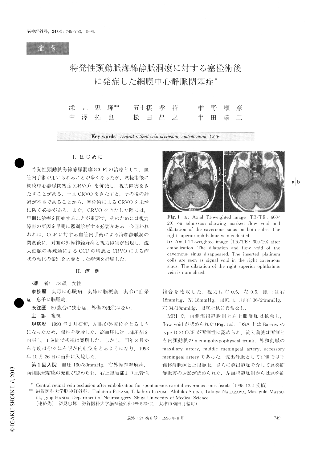
特発性頸動脈海綿静脈洞瘻(CCF)の治療として,血管内手術が用いられることが多くなったが,塞栓術後に網膜中心静脈閉塞症(CRVO)を併発し,視力障害をきたすことがある.一旦CRVOをきたすと,その後の経過が不良であることから,塞栓術によるCRVOを未然に防ぐ必要がある.また,CRVOをきたした際には,早期に治療を開始することが重要で,そのためには視力障害の原因を早期に鑑別診断する必要がある.今回われわれは,CCFに対する血管内手術による海綿静脈洞の閉塞後に,対側の外転神経麻痺と視力障害が出現し,流入動脈の再疎通によるCCFの増悪とCRVOによる症状の悪化の鑑別を必要とした症例を経験した.
A patient with spontaneous carotid-cavernous sinus fistula, who developed central retinal vein occlusion (CRVO) after interventional surgery, was reported.
A 78-year-old woman was admitted with symptoms of right abducens palsy and conjunctival injection. Digital subtraction angiography (DSA) revealed bilater-al carotid cavernous sinus fistulas supplied by duralbranches of the carotid arteries.
After embolization of the feeding arteries and the right cavernous sinus using PVA and platinum coils re-spectively, her symptom improved gradually. Six months after the embolization, she was readmitted be-cause of blurred vision and abducens palsy on the left side. Engorgements of the cavernous sinuses and drain-ing veins, and the shunt flow of the fistula were muchless on DSA than those seen previously, but ophthal-mologic studies showed an impending central retinal vein occlusion (CRVO) in her left eye.
Prognosis of CRVO is generally poor. We discussed the mechanism of CRVO occurring after interventional surgery for CCF and the tactics for preventing it, and early detection of its development.

Copyright © 1996, Igaku-Shoin Ltd. All rights reserved.


